


▶ Previous Artlcle : #2-1. Anatomic Study of Skin Surface II
F. Facial muscles
Facial muscles are divided into two subunits; muscles involved in facial expressions and those involved in mastication (Figure 2). Close familiarity with expression muscles is required for aesthetic procedures. These muscles are innervated by facial nerves. Unlike limb muscles that attach to the bone, facial expression muscles are attached to the skin and muscles. With this unique characteristic, they produce various facial expression through muscle movement. Among muscles around the eyes (Figure 3), the corrugator supercilii muscle, depressor supercilii muscle, medial head of the orbital portion of orbicularis oculi muscle, and procerus muscle are involved in creating glabella wrinkles. The corrugator supercilii muscle is divided into transverse head and oblique head which have differing direction of actions due to their divergent origins and insertions. The depressor supercilii muscle and oblique head of corrugator supercilii muscle originate from around the inner orbit and run parallel to each other to insert into the medial eyebrow dermis. The medial head of the orbital portion of orbicularis oculi muscle originates from the medial canthal ligament and inserts into the medial eyebrow dermis. Considering the origin and insertion, the oblique head of the corrugator supercilii muscle, depressor supercilii muscle, and medial head of the orbital portion of orbicularis oculi muscle pull the medial end of eyebrow downward. The transverse head of corrugator supercilii muscle originates from the superomedial orbital margin and inserts into the dermis at the middle third of the eyebrow. This muscle draws the eyebrow inward. Anatomical understanding of the eye area is used in various treatments of glabella wrinkles.
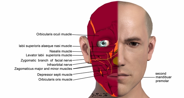
Figure 2.
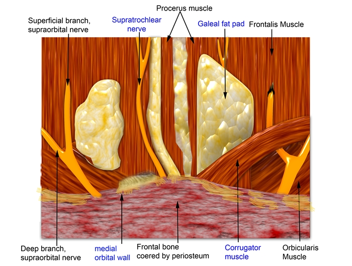
Figure 3.

[Advertisement] Selene(Diode hair removal Laser) – Manufacturer: (www.senbitec.com)]
G. Lymphatic system
The facial lymphatic system is similar to the venous circulation but also differs from it in many ways. The lymph vessels circulate into the same side of the system and course in the direction from surface to deeper areas, medial to lateral and top to down.
The lymph node system of the face is called pericervical node group that form the first filter system at the craniocervical border. From the front, the pericervical node group consists of the submental node (two thirds into the middle of the chin), submandibular node [nose, medial cheek (including medial eyelid), upper lip], parotid node [lateral face, forehead, ant. scalp (frontotemporal), lateral eyelid], retroauricular node (parietal scal) and occipital node (occipital scalp), etc. The mid lower lip can be drained by ipsilateral and contralateral submental and submendibular nodes. The lymph nodes of the face drain into the internal jugular chain of the neck after systemic circulation of the face.
The cervical lymphatic drainage is largely divided into superficial node system and deep node system (internal jugular nodes, spinal accessory nodes and transverse cervical nodes). The internal jugular node is the final pathway that receives all drainages from the head and neck. Located inferior to the SCM, it can be easily palpated by holding down on the muscle and turning the head in the ipsilateral direction. Skin cancer of the head and neck spreads through this channel and diagnosis of lymph node enlargement often precedes diagnosis of cancer. However, lymph drainage varies widely between individuals and lymph node enlargement is found in about 20~50% of healthy population.
-To be continued-
▶ Next Artlcle : #2-3. Anatomic Study of Skin Surface II


