▶ Previous Artlcle : #2-1. Functions of the Venous System
Capacitance Vessel Function
This function refers to the role of veins to store most of the blood volume in the body. About 80% of the entire blood volume is stored in the venous system such as the venous vessels, right atrium and ventricle, pulmonary veins/arteries and left ventricle, etc. Two thirds of the entire blood volume is held in the venous vessels from venules to large veins. Most of this blood volume is stored in the deep venous system. Unlike arterial vessels, veins have thin, pliable walls which allow them to flexibly store large volumes of venous blood.
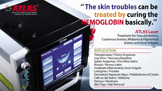
[Advertisement] ATLAS(Long pulse 532nm & 940nm dual wavelength) – Manufacturer: MEDRO(www.medro.net)
Musculovenous Pump Function
Due to muscle contraction in lower limbs, 90% of venous blood is carried into the heart through deep veins. Muscle contraction in lower limbs pump the blood in deep veins upward to the heart. During muscle contraction, the valves within perforating veins block the blood flow toward superficial veins. On the other hand, during muscle relaxation, deep venous pressure is lower than the superficial venous pressure, which sends superficial venous blood into deep veins through perforating veins. This mechanism of called the calf muscle pump or peripheral heart.
Among the calf muscles, the soleus muscle carries out the primary pumping function. Walking forces this muscle to pump blood to the heart through deep veins. As the calf muscles play an important role of the peripheral heart, gastrocnemius muscle resection or neurectomy in an aesthetic calf reduction surgery drastically weakens this peripheral heart. In terms of hemodynamics, this procedure is not desirable as it inhibits the return of venous blood to the heart.
Most of the lower limb venous blood is pumped into the heart by the above-mentioned mechanism. However, 50% of perforating veins in the foot lack valves which may cause reverse flow of blood into the epidermis. Due to this anatomical factor, sclerotherapy with injection of a potent sclerosing solution in the foot can create deep veins thrombosis and thus requires great caution.
Lower limb veins are equipped with valves (especially deep veins) and these veins are embedded in muscles that work as blood pumps. Calf muscles surrounded in strong fascia can elevate the intramuscular pressure up to 250mmHg during contraction. Lower limb muscles go through repeated contraction-relaxation and push the blood in the veins upward to the heart. The musculovenous pump works as a peripheral heart, continuously lowers the blood flow into lower limbs and prevent accumulation of tissue liquid or tissue edema that may occur due to rising venous pressure in the standing position.
When this lower limb pump is impaired, various abnormalities occur. Contraction of each muscle squeezes the deep veins and thereby push the blood upward. Venous valves prevent reverse flow of the blood during muscle relaxation. When the venous blood is not pumped upward, the distribution of nutrients and oxygen occurs in peripheral capillaries.
When this function is impaired, waste accumulates in the skin or soft tissues, causing various skin conditions. This may result in noticeable talengiectasia on the inner sole of the foot. When this condition exacerbates, it could progress into dermatitis which is often recalcitrant to treatment. Such changes in the microcirculatory system of the lower limb indicate the onset of venous disorder. If left untreated, it could result in skin necrosis.
Mechanism of Musculovenous Pump
During muscle relaxation, the vein and venous sinus are filled with blood.
Muscle contraction compresses veins, and blood within the venous sinus is pushed toward the heart.
Normal valves prevent the reverse flow of blood.
When the muscle is relaxed, veins are dilated and filled again with venous blood.
Hemodynamics of Varicose Veins
Blood sent to the lower limb through arteries return to the heart due to the actions of calf muscles (gastrocnemius) and blood vascular valves. As mentioned above, in normal hemodynamics of the lower limbs, muscle contraction opens the valves and pushes venous blood into large veins. The lower limb muscles are called calf muscle pump or peripheral heart.
Valves within the veins are generally located where venous tributaries converge. The valves are arranged so that the venous blood in lower limbs flow from upward and outward. Valves grow in number toward the lower areas.
In case of varicose veins arising from dysfunction of venous valves, blood flow is reversed and deep venous blood flows into superficial veins during muscle relaxation. Under normal circumstances, during muscle relaxation, deep venous pressure falls below the superficial venous pressure which allows the influx of superficial venous blood into the deep veins through perforating veins. Varicose veins occurs when this blood flow is reversed.
A major cause of varicose veins is venous valve insufficiency. Blood regurgitation due to valve insufficiency raises the venous pressure and vein diameter resulting in turbulent flow of the blood leaked through the valves. When this phenomenon persists, saccular dilation of venous walls occurs due to the turbulent flow below the valves. When this condition persists, it could lead to fibrosis and long-term venous hypertension worsens valve insufficiency.
The saccular dilation or turbulent flow can be detected by ultrasonography or surgery. Hereditary venous wall or valve deficiency is another etiological factor. During an ultrasound examination, the sound of turbulent flow is larger and longer-lasting in patients with longer duration of varicose veins, thicker veins and saccular dilation. The sound of regurgitated blood is also louder and longer in patients with severe varicose veins, although this is not always the case. The regurgitation sound of varicose veins originating from GSV (great saphenous vein) is also larger and longer compared to that of SSV(small saphenous vein) with the same thickness.
-To be continued-
▶ Next Artlcle : #3-1. Anatomy of lower limb veins


