▶ Previous Artlcle: #6-4. Dermal melanocytic lesions
Post-inflammatory hyperpigmentation
Post-inflammatory hyperpigmentation may occur after procedures such as chemical peel, laser, dermabrasion, and liposuction.
It can also occur as a complication after trauma or after inflammatory acne or dermatitis.Histologically, melanin is increased in the epidermis andmelanophage is increased in the upper dermis and around the blood vessels.
It usually improves over time, but if the patient wants a quick improvement for cosmetic purposes, it can be improved a little faster with lasers, peeling, and whitening products.
In particular, the combined use of 1064 pico-second laser,long pulsed Alexandrite laser, and Er:YAG laser can effectively improve it without sequelae.
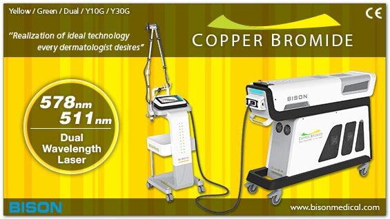
[Ad. ▶ COPPER BROMID(Yellow/Green Laser) – Manufacturer: BISON(www.bisonmedical.com)]
Seborrheic keratosis
Seborrheic keratosis is a benign tumor lesion that is most commonly seen in the elderly. The causehas not been clearly known, and it occurs as epithelial cells proliferate.
At first, it begins with a light brown spot with a clear borderline and grows larger and thicker, developing into a lesion with a rough, cracked surface like the wart surface, or it progresses to a dome-shaped lesion with horn cysts.Most cases can be diagnosed by clinical findings.
Histological findings includepapillomatosis, acanthosis, and hyperkeratosisin the epidermis.The base of the lesion progresses alongside the epidermal dermal border.It can be treated with Cryotherapy, CO2 laser, Er-YAG laser, Q-switched Ruby laser, and long pulsed Alexandrite laser.
- To be continued


