


▶ Previous Artlcle: #6-1. A Case of Treatment for a Side Effect of Facial Liposuction
Cheeks do not have a lot of subcutaneous fat and consist of superficial muscular aponeurotic system (SMAS) as a single layer deep within the skin.
Even when tumescent solution is injected, the required dose for each cheek is small at 10 to 20cc.
After injection, a 10 minute wait is needed until the effects of pinephrine fully appear before fat sucking.
In addition, cheek liposuction necessarily requires the use of a 18-gauge thin cannula. It is contraindicated to use piston pressure of 10cc or above.
What matters is to suck fat out as if slowly sliding the cannula out while using a low piston pressure of about 5cc.
The hole of cannula must not be approached in the direction of skin, and fat must be sucked in side and downward directions.
[Advertisement] MAGNUM(Q-switched Nd:YAG Laser) – Manufacturer: (www.i-dana.com)]
Prevention against Side Effects of Cheek Liposuction
The most common reason for skin dimpling after cheek liposuction is an excessive tissue adhesion between skin and muscle fascia, which occurs due to excessive fat sucking.
To prevent this side effect and achieve a satisfactory result, only yellow-colored fat must be sucked out while necessarily leaving a fat layer as it is.
It is also important to avoid the formation of a lot of tissue adhesions by using local anesthesia and having the cannula approached in the direction of muscle fascia, the area from which patients complain of pain, for fat sucking.
A patient who is introduced in this issue to present a relative treatment case is a woman aged 30 years who underwent cheek liposuction at a domestic hospital specializing in liposuction.
However, pain had been severe since the end of the procedure, and bruising lasted more than a month.
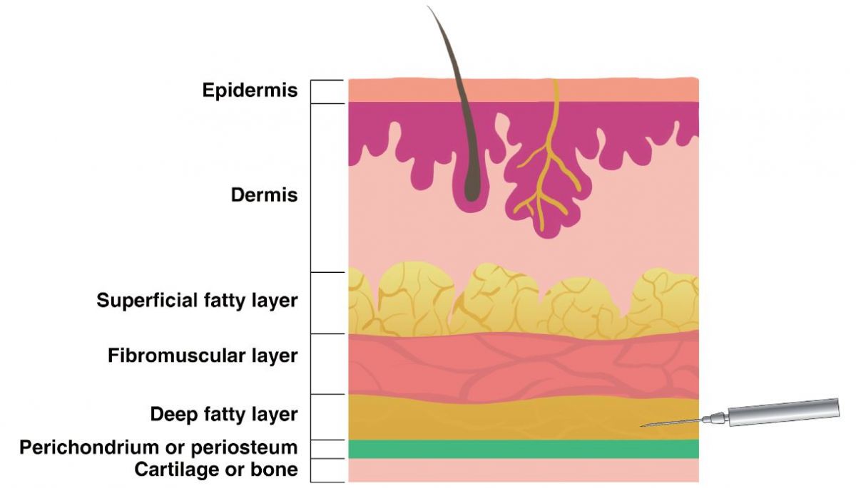
Figure 1. Soft tissue layer, SMAS(Fibromuscular layer).
-To be continued


