


▶ Previous Artlcle : #18-4. Picosecond Laser
As a high-intensity laser beam is irradiated to the tissue and the photons are absorbed into melanin, melanin that absorbed the photons begin to emit electrons, causing a flood of free electrons.
The rapid increase in free electrons enhances the electron plasma density, forming hot plasma balls and diffusing the heat into the surrounding tissue to form vacuoles within the tissue (see Figure 5).
Whether the plasma is formed in the dermis or the epidermis can determine the effects of tissue regeneration.
.jpg)
[Advertisement] FCR® (Fractional Prickle CoralCalcium Regentron) – Manufacturer: (www.illglobal.com)]
Thus, the operator has to consider where to form the plasma to improve treatment of the legion more accurately.
In normal laser therapies, the higher the fluence, the deeper the penetration into the tissue.
However, the opposite is true in case of a picosecond fractional laser, owing to the plasma shield effect.
A plasma shield indicates a formation of plasma when a when a high intensity laser beam is focused on the tissue and the LIOB occurs on the spot.
The gas molecules caused by the formed plasma inhibit the photons from penetrating deeper, and the photons accumulate from the periphery to the rear, or toward the epidermis, forming plasma balls on the epidermis or near the epidermis to creating vacuoles.
The higher the fluence, the more plasma is formed on the epidermis.
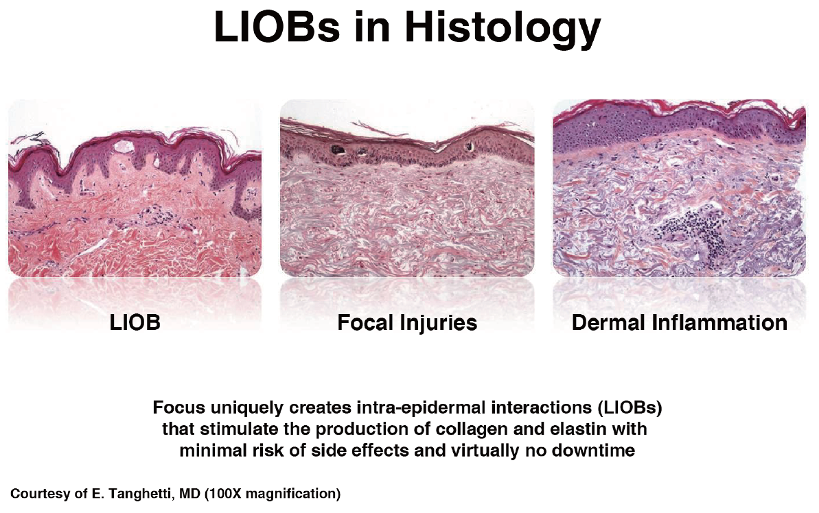
Figure 5. Process of LIOB.
-To be continued


