


Surgical Treatment of Hypertrophic Scar and Keloid
Surgery is not the first choice for hypertrophic scar or keloid. In most case, injection therapy or other conservative treatments are first-line treatments, because of high recurrence and exacerbation after surgery. Postoperative recurrence and exacerbation depend on several factors, including the size and site of lesions, patient’s age and sex. Commonly, hypertrophic scar or keloid, in young patients, extremities and pre-sternal area, easily recurred or exacerbated.
Surgical methods for hypertrophic scar or keloid can be roughly divided to complete excision, intralesional partial excision and core excision. Complete excision is not recommended, except for particular cases, while core excision is a useful method for globular lesion, as in the case of keloid in the ear.
It is very hard to cure a hypertrophic scar or keloid by surgical treatment alone, and it has to be combined with various postoperative conservative treatments. On the assumption that combination is continued, surgery can be considered as the first-line therapy for large lesions that can be hard to obtain immediate effect by conventional treatment.
[Advertisement] SKIN THE NEXT – Manufacturer: NEXGEN(www.nexgenbiotech.com)
1. Complete excision
Complete excision can be applied to hypertrophic scars, but is not recommended for keloid due to the risk of exacerbation. Conventionally, a number of literature have reported high cure rate by complete excision followed by radiotherapy, both of which are hardly comfortable for patients.
Complete excision typically uses simple incision and suture, followed by skin graft, which is not recommendable for keloid skin due to the possibility of creating a new keloid.
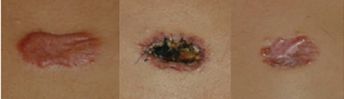
Figure 1. Complete excision (graft) of keloid
2. Intralesional partial excision
This is the surgical technique most commonly used for scar and keloid. The primary aim is to reduce the volume, not removal, of the lesion to facilitate injection therapy and others.
Intralesional partial excision is associated with few recurrence or exacerbation for unknown reason, but possibly because keloid tissue works as a barrier preventing intralesional exacerbation. Personally I also think that intralesional excision could be a good choice, if combined with proper postoperative treatments,[reference; Kim et al ‘Study on Intralesional excision of Keloid’ published in the Korean Journal of Dermatology in 2009].
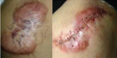
Figure 2. Intralesional partial excision for a large keloid on the shoulder
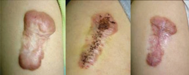
Figure 3. Supportive therapy (steroid injection) after intralesional partial excision
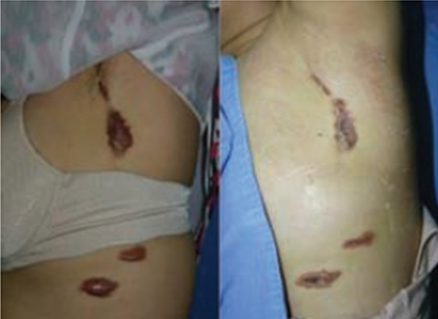
Figure 4. In multiple large lesions, intralesional partial excision can reduce the volume and number of postoperative steroid injection.
3. Core excision
For keloid in the ear, the outer skin can be dissected to make a thin flap, which can be covered again after near complete excision of the keloid. This technique is often called a core excision, but is technically an intralesional excision. There are a lot of reports about very good clinical results after applying this method to keloids in the ear.
My experiences also suggest that this surgical technique wound be the best choice for keloid in the ear, if combined with continuous supportive therapies.
There are a variety of supportive therapies available after core excision. Special assist device, such as pressure earring, can be applied for lesions in the ear. Topical imiquimod was also reported recently as providing a good result.
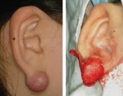
Figure 5. Core excision: the outer skin was dissected and the solid lesion inside was removed.
- To be continued-
▶ Previous Artlcle : #7. Surgical Treatment of Scar Ⅰ
▶ After Artlcle : #9-1. Case series of Scar Treatment


