▶ Previous Artlcle : #3-1. Ultrasound Cavitation
In 1990, Child et al. reported lung hemorrhage in mice exposed to 1-MHz ultrasound under the threshold pressure of 2 MPa for 1 μm pulse; the hemorrhage was caused by the destruction of lung cells and capillary endothelial cells due to the inertial cavitation or transient cavitation. Such damages can be more severe with higher peak pressure, while attenuated with higher frequency.
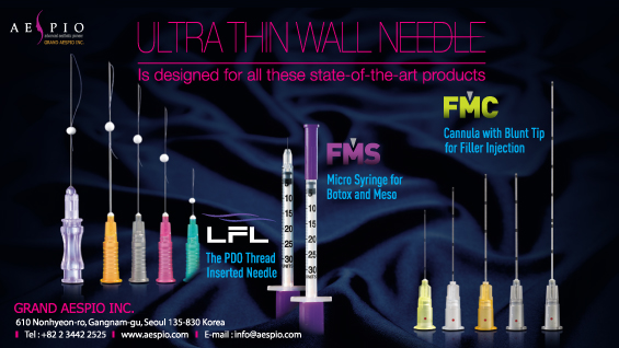
[Advertisement] ULTRA THIN WALL NEEDLE – Manufacturer: AESPIO(www.aespio.com)
However, whether the cavitation is the sole cause of pulmonary hemorrhage induced by ultrasound is controversial. As alveoli are exposed to air bilaterally and the alveolar wall is a membrane covered with water and surfactant, the acoustic impedance can be abruptly increased when ultrasound passes through the wall, possibly causing tissue damage. In case of intestines, however, inertial cavitation is commonly agreed as the main cause of hemorrhage by diagnostic ultrasound devices. In 2000, Miller and Gies et al. reported that intestinal hemorrhage was increased when artificial microbubbles were injected systematically as a contrast agent for diagnosis. Intestinal hemorrhage was more common at a lower frequency (0.7 MHz) than higher one (3.6 MHz).
Cavitation induces histological changes in the skin as well. Sonophoresis is a phenomenon that occurs with increased permeability of skin tissues or cells by ultrasound. With a 15-μm barrier composed of corneum and lipids, the skin have a very low permeability to foreign substances in natural conditions. Therefore, sonophoresis has been used for the percutaneous penetration of desired substances into the circulation.
In 1989, Levy et al. reported that transdermal permeability to mannitol was 5 to 20 times greater for 1-2 hours after 5 minutes of irradiation of 1 and 3 MHz ultrasound with continuous wave at 1.5 W/cm². Merino et al. (2003) reported that the permeability to more various sizes of substances was further enhanced when a lower frequency ultrasound was used.
To explain the mechanism of this phenomenon, Tchibana et al. (1992) described that microbubbles in cavities filled with a liquid, such as sweat glands and ducts of sweat glands, acted as nuclei for cavitation in the corneum. The authors concluded that the expansion and explosion of the microbubbles created cracks in the lipid bilayer, enhancing the permeability. On the contrary, Tezel et al. (2003) contradicted this mechanism by microbubbles in the corneum. They insisted that microbubbles located close to the surface outside the epidermis would work as cavitation nuclei, stating that the diameter of bubbles observed during ultrasound irradiation was too large compared to 15-μm-thick corneum. They proposed 3 possible mechanisms of the crack formation in the lipid bilayer by cavitation: the first was the diffusion of shock wave at the time of symmetric collapse of microbubbles. Pecha et al. (2000) have already reported that bubble explosion generates shock waves, which are propagated to several micrometers from the bubbles at the speed of 4000 m/s. The bunch of shock waves made from the collapse of numerous microbubbles are collectively called a ‘cavitation noise.’ The difference in pressure generated in the course of propagation of the bunch of shock waves through 15-μm corneum is as big as 100 MPa. This pressure may be the cause of the enhanced skin permeability. The second was that the penetration of acoustic microjet into the corneum may generate the crack. Asymmetric collapse of microbubbles occured close to a barrier generates a liquid jet to one direction, which is called a ‘water hammer.’ The pressure of this water hammer is strong enough (several kilobars) to penetrate the barrier. The third is the accumulated pressure, describing that even a low instantaneous pressure is enough to penetrate the barrier when exposed for a long time.
The mechanism of enhanced transdermal permeability by ultrasound is not clear yet, but these 3 mechanisms proposed by Tezel et al. are acknowledged as relatively acceptable hypotheses. Sonophoresis deserves another article to discuss about. Cavitation is also used for thrombolysis. Ultrasound is used to enhance the infiltration of thrombolytic agents or to dissolve thrombus directly. Blinc et al. (1993) found that 1- and 2.2-MHz high-intensity ultrasound increased fibrinolysis. The authors reported that, as in the skin, cavitation enhanced the permeability of thrombolytic agents into thrombus, resulting in further increase in fibrinolysis. Atar et al. (2004) demonstrated that in vivo and in vitro thrombolytic effect can be induced by cavitation and jet flow occuring close to the wall of thrombus, even without a thrombolytic agent.
Cavitation induced by ultrasound in blood vessels, especially in the vessel wall, also increases extravasation. In the cerebral blood flow barrier, low-intensity ultrasound makes contrast agents continuously oscillated around the wall, which generates shearing forces and microstreaming. Higher-intensity ultrasound makes the oscillation stronger, ultimately resulting in explosion.
In this process, sonoporation of the capillaries occurs, facilitating the delivery of drugs or genes. The ultrasound contrast agents, mentioned in this article, are artificial encapsulated microbubbles, which have been intravascularly injected to increase the scattering of ultrasound and to obtain more clear vascular images. Recently, they are also used as a medium for gene or drug delivery for its cell membrane perforation effect.
-To be continued-
▶ Next Artlcle : #4-1. Ultrasound-Mediated Adjunct Therapy


