▶ Previous Artlcle : #12-1. Tissue Repair Stimulating Agent with Nucleic Acid
As explained in the article, PDRN has been already approved as an injection in PRP or stromal vascular fraction(SVF) treatments and does not require the cumbersome process of blood collection, isolation and purification. Another advantage of PDRN is that there is no variation for individual patients. To my knowledge, however, there still seems no objective and prospective clinical study on the difference in clinical efficacy compared to the existing PRP or SVF. Additional studies are needed to provide more objective scientific data to improve its clinical usage.
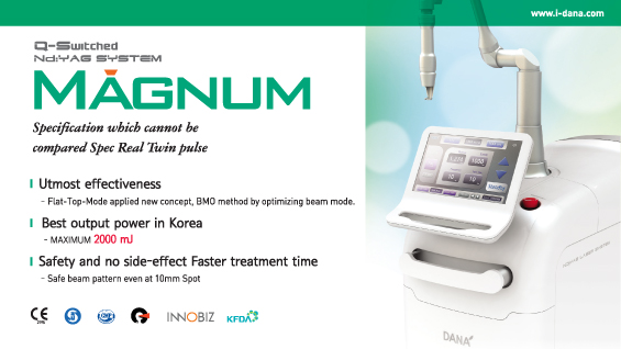
[Advertisement] MAGNUM(Q-switched Nd:YAG Laser) – Manufacturer: (www.i-dana.com)]
As hinted in the brand name ‘Placentex’, PDRN was characterized at first by its wound healing activity by facilitating the tissue repair process in human placenta. However, it is now produced by extraction from trout sperm, which is why PDRN injection is referred to as ‘trout injection’ at some Korean hospitals.
As briefly mentioned above, PDRN is comprised of a part of low molecular DNA which is the active ingredient in an agent used for wound healing and skin regeneration. It consists of deoxyribonucleotide polymers of 50-2,000 base pairs where purines and pyrimidine nucleotides are combined by a phosphodiester bond, providing purines, pyrimidines as well as deoxyribonucleotides and deoxyribonucleosides. PDRN mostly affects nucleotides. Nucleotides and nucleosides are known to facilitate the growth of various cells, including fibroblasts, chondrocytes and osteoblasts, and to accelerate nucleic acid synthesis and wound healing, probably by facilitating the salvage pathway that produces nucleic acid with lower energy consumption and activating adenosine A2A receptor. PDRN is also effective in skin regeneration, chronic wound, bedsore and burn. Among the three phases of wound healing process, the proliferative phase and remodeling phase seemed to be influenced by PDRN the most. However, recent reports suggest that PDRN might affect the early inflammation phase and the whole wound healing process to stimulate wound healing.
There is relatively large volume of literature on the wound healing effect of PDRN in animal models. In a test that compared the effect of PDRN on a diabetic wound in diabetic mice, it was suggested that PDRN increased VEGF message and protein wound content and that its selective activity was effective for diabetic wound healing. When compared to wet dressing with saline solution in mice with deep-dermal s second degree burns, PDRN increased epithelial proliferation and extracellular matrix maturation, facilitating wound healing, increasing angiogenesis and reducing the time required for wound healing.
In a clinical study comparing the wound healing effect on the donor site of skin graft, PDRN stimulated reepithelialization when the topical cream was applied directly to the donor site. Wound healing was also improved with intramuscular and subcutaneous injection to the surrounding area.
PDRN was also reported to have an influence on fibroblasts, which play important roles in collagen synthesis in wound healing. PDRN facilitates the growth of cultured human skin fibroblasts, promotes production of collagenic and noncollagenic matrix proteins, and prevents scars through normal matrix formation. Below is a summary of my study on the wound healing effect of PDRN:
Existing mouse wound healing models mostly involve wound contraction, not epithelialization. We used a silicon ring around full-thickness wounds so that the ring could work as a splint (support) and minimize contraction. The mechanism of PDRN was investigated in a modified mouse wound model where granulation tissue formation and reepithelialization contributed to wound healing in manners similar to the human wound healing process.
Forty-two white male mice (age, 7 weeks; weight, 30-40g) were randomized to one of three groups. A circular, full-thickness skin defect wound (10×10mm) was made on the back of the mice, and the surrounding area was fixed with a silicon ring splint. Group 1 received intraperitoneal PDRN 8mg/kg/day for 12 days, Group 2 received direct administration to the wound, and Group 3 received intraperitoneal saline solution. For histological analysis, 4 or 5 mice per group were sacrificed at Day 4, 8 and 12, and the wound tissue was collected for H&E staining. The effect of PDRN on wound healing was evaluated by macroscopic change in wound size (degree of epithelialization) and the extent of granulation tissue formation. Immunohistochemical staining and Western blotting using VEGF, transforming growth factor beta (TGF-β) and CD31 were used to quantify the expression of angiogenesis, the number of blood vessels, and growth factor increase. Fibroblasts that were deemed to be associated with the mechanism of wound healing were cultured and the change in the cell number depending on the concentration of PDRN (100-2,000μg/mL) was observed to determine an appropriate concentration.
The results showed that PDRN stimulated wound reepithelialization and granulation tissue formation, increased microvessel density, stimulated the expression of VEGF, TGF-β and CD31, and also facilitated angiogenesis. An appropriate concentration of PDRN (100-1,000μg/mL) stimulated the proliferation of fibroblasts, and the maximum effect was shown at the concentration of 1,000μg/mL.
In conclusion, this modified animal model showed that PDRN facilitates wound reepithelialization and granulation tissue formation by stimulating the expression of VEGF, TGF-β and CD31 as well as the differentiation and maturation of fibroblasts. The main mechanism of action of PDRN could be roughly summarized as anti-inflammation, cell proliferation and stimulation of regeneration factors.
By facilitating the wound healing process, rapid tissue regeneration and differentiation of different cell types and by improving microvascular circulation and anti-inflammatory activity through the expression of VEGF, PDRN can be utilized in various clinical fields from wound healing to tissue regeneration, scar treatment, wrinkles, skin aging, photoaging, skin elasticity improvement and alopecia treatment as well as for the treatment of chronic pain and degenerative arthritis. It is expected that the application of PDRN will only broaden. Further studies including systematic clinical studies are needed in the future to support its efficacy.
References
❶ Tonello G, Daglio M, Zaccarelli N, et al. Characterization and quantitation of the active polynucleotide fraction (PDRN) from human placenta, a tissue repair stimulating agent. J Pharm Biomed Anal 1996; 14: 1555-60. ❷ Galeano M, Bitto A, Altavilla D, et al. Polydeoxyribonucleotide stimulates angiogenesis and wound healing in the genetically diabetic mouse. Wound Repair Regen 2008; 16: 208-17. ❸ Bitto A, Polito F, Altavilla D, et al.Polydeoxyribonucleotide (PDRN) restores blood flow in an experimental model of peripheral artery occlusive disease. J Vasc Surg 2008; 48: 1292-300. ❹ De Aloe G, Rubegni P, Biagioli M, et al. Skin graft donor site and use of polydeoxyribonucleotide as a treatment for skin regeneration: a randomized, controlled, double-blind, clinical trial. Wounds 2004; 16: 258-63. ❺ Altavilla D, Bitto A, Polito F, et al. Polydeoxyribonucleotide(PDRN): a safe approach to induce therapeutic angiogenesis in peripheral artery occlusive disease and in diabetic foot ulcers. Cardiovasc Hematol Agents Med Chem 2009; 7: 313-21.
-To be continued-
▶ Next Artlcle : #13-1. Biologically Derived Collagen: Elastin Dermal Substitute


