


This article will discuss HUVECs (human umbilical vein endothelial cells). HUVECs are cells isolated from the umbilical vein. They are also isolated from the cord blood and extensively used in the research on modulating the endothelium function.
The umbilical cord blood is the blood that remains in the tissues of the umbilical cord. It is rich in hematopoietic stem cells and serves as a reservoir for stem cells. It is used along with the bone marrow and peripheral blood for mobilizing stem cells. Cord blood is an efficient source of stem cells as its provider feels little burden. HUVECs can be obtained from the cord blood, and this provides many advantages to research.

[Advertisement] Selene(Diode hair removal Laser) – Manufacturer: (www.senbitec.com)]
HUVECs can be isolated by treating the umbilical vein with collagenase before culture. After isolation, the cells are should be confirmed if they have derived from the endothelium using von Willebrand factor (vWF, expressed in vascular endothelial cells and helps platelet aggregation) antibodies. HUVECs isolated and cultured this way facilitate in vitro studies of cells that are difficult to observe in vivo. HUVECs are endothelial cells that contain hematopoietic stem cells. Therefore, they are particularly useful for identifying the function and role of endothelial cells.
Endothelial cells are superficial squamous cells that cover the inner walls of the blood vessels and lymphatic ducts. They belong to the group of cells that make up blood vessels. Vascular endothelial cells are involved in maintaining homeostasis. The vascular endothelium maintains anticoagulatory and anti-inflammatory barriers between the blood and tissues. Thanks to these functions of the vascular endothelium, the human body can adequately respond to infiltration of various pathogens.
Endothelial cells have following functions in the human body.
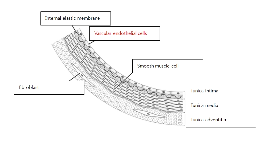
Image 1. Diagram of the endothelium.
-To be continued-


