


▶ Previous Artlcle : #1-2. Anatomic Study of Skin Surface I
The next zygomatic branch and buccal branch has many smaller branches and lie deep in the tissues. Damage is not likely but even when damaged, motor impairment is rare. The fourth branch, marginal mandibular branch lies shallow and is susceptible to damage as it runs in one nerve fiber. This branch innervates the orbicularis oris and the depresser labi muscle group around the mouth. When damaged, the mouth twists upward on the undamaged side. The last branch, cervical branch innervates the plastysma and has little clinical significance when damaged.

[Advertisement] Selene(Diode hair removal Laser) – Manufacturer: (www.senbitec.com)]
One of the most severe complications of a facial surgery is nerve damage. Most facial nerve branches run below the SMAS and are protected by the facial muscles or fascia, making damage minimal. However, the temporal branch and mandibular branch are not connected to other branches and are often damaged in the lateral, posterior face.
The temporal branch of the facial nerve is most often damaged in dermatological surgeries. It is damaged between the ear and lateral canthus, in other words, around the zygomatic arch. As the protective SMAS is interrupted in this area, the nerve lies in the subcutaneous fat. Especially, in an elderly patient with hollow cheeks and very little fat layer, the SMAS can be incised unwittingly leading to nerve damage. Damage prone areas can be determined in two methods (Figure 10, 11). First, the triangle connecting three points – the lateral most points of the earlobe and eyebrow and the lateral most point of the highest forehead line – is a damage prone zone. Second, the nerve lies between 1cm inferior to tragus and 1 cm lateral to eyebrow.
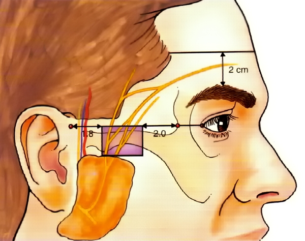
Figure 10. temporal branch of facial nerve index (A).
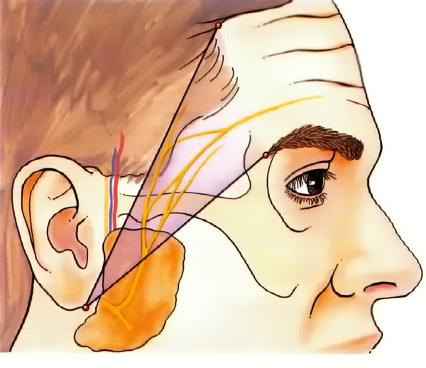
Figure 11. temporal branch of facial nerve index (B).
Next is the mandibular branch. Posterior to the area where the anterior-inferior masseter and inferior mandible meet (facial artery intersection), it runs from 1~2cm below the inferior mandible to cross this point along the inferior mandibie. This course makes damage very likely. Moreover, around the age of five (or around puberty in some cases) the mastoid process is inadequately developed, which exposes facial nerve to damage as only thin subcutaneous fat surrounds it.
-To be continued-
▶ Next Artlcle : #2-1. Anatomic Study of Skin Surface II


