


▶ Previous Artlcle : #1-3. Anatomic Study of Skin Surface I
In the broad topic of skin anatomy, the previous article discussed anatomical terminology of the skin, superficial musculoaponeurotic system (SMAS), and facial nerves, etc. This article will take a look at facial sensory nerves, facial vascular anatomy, facial muscles and lymphatic system, etc.
D. Facial sensory nerves
The sensory nerves in the head and neck are divided into two areas; 1) facial and cranial nerves and 2) the neck nerves. The face and anterior scalp are innervated by three branches of the tigeminal nerve. More specifically, the ophthalmic, maxillary, and mandibular divisions innervate posterior cranium and the inferior boundary of the lower jaw, and cervical nerves (C2-C4) innervate the neck (Figure 1). In dermatologic surgery, familiarity with the sensory nerves is important not only for prevention of post-procedure damage but more for safer procedure with appropriate local anesthetics and successful outcome.
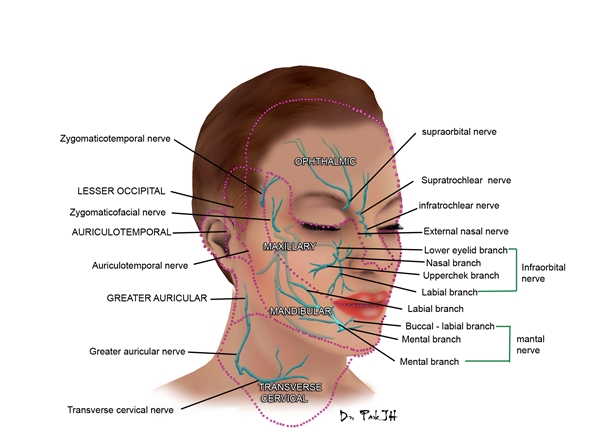
<Figure 1>
The ophthalmic branch of tigeminal nerve innervates the forehead, anterior cranium, nose, upper eyelid, and conjunctiva through the supratrochlear, supraorbital nerves. The maxillary branch innervates the cheek, lateral face, lower eyelid, lateral nose, and upper lip, etc. through the zygomaticofacial, zygomaticotemporal, infraorbital nerves. The lowest mandibular branch innervates the lower lip, cheek, temple and anterior two thirds of tongue, mouth floor, and mandibular teeth through four important branches (including mental nerve); the inferior alveolar nerve, lingual nerve, buccal nerve, and auricular nerve.
The depth of the neural course lies between subcutaneous fat and SMAS and therefore, is easily exposed during surgery, but most cases of damage recover in time.

[Advertisement] FCR® (Fractional Prickle CoralCalcium Regentron) – Manufacturer: (www.thermoceutical.asia)]
E. Facial vascular anatomy
Most of the face is vascularized by branches of external carotid artery, a section of common carotid artery. However, the midface, the area around the eyes, superior two thirds of the nose and mid forehead are mainly vascularized by the ophthalmic artery, a branch of internal carotid system. Additionally, the intersection of facial artery and superficial temporal artery, branches of external carotid artery provide blood to this area. The facial artery has high pressure and hemostasis often requires ligation of both ends as well as simple electrocautery. Due to many intersections, vascularization is maintained despite disruption of one artery and therefore, does not require anastomosis. Facial veins mostly lie parallel to the arteries. As the facial veins lack valves, a characteristic venous structure, extracranial infection can be intracranially transmitted without any inhibition. Infection around the nose particularly requires great caution.
-To be continued-
▶ Next Artlcle : #2-2. Anatomic Study of Skin Surface II


