


Supratrochlear Artery
The most serious complication of injectable dermal fillers is visual loss, which is most common with injections in the glabella. Data on the frequency of visual loss at this site are consistent in domestic and foreign literature. The supratrochlear artery is often involved and we need to be clearly aware of the course of the supratrochlear artery to avoid it during injection. However, controversy still exists over exactly where the supratrochlear artery perforates the frontalis muscle for its superficial emergence or which structures it lies above. Pessa named the crease in the lateral glabella as ‘corrugator crease’ and argued that this crease coincides with the course of the supratrochlear artery. He also argued that the artery passes below the frontalis muscle and corrugator supercilii muscle.
On the other hand, Ugur et al. reported that the supratrochlear artery courses around the medial canthal line, which lies lateral to the glabellar frown line (corrugator crease) in his doppler imaging and cadaver study. Reece et al. argued that the supratrochlear artery passes along the periosteum to exit the medial orbit and divides into a superficial and deep branch. The deep branch rises along the periosteum but the superficial branch perforates the frontalis muscle 1.5mm above the supraorbital rim and runs in the subcutaneous layer.
Based on these various reports, I have concluded that the course of the supratrochlear artery mostly coincides with the corrugator crease and the deep branch does exist but the superficial branch is a thicker main branch. I believe the superficial branch perforates the frontalis muscle slightly above the supraorbital rim. However, there are variations to the location of these structures among patients and glabella injections should be carried out with great caution.

[Advertisement] Ultra Skin/Pastelle – Manufacturer: WONTECH(www.wtlaser.com)
Forehead Fat Compartments
The fat compartment of the forehead only has superficial fat above the frontalis muscle and lacks deep fat. The forehead is divided into the central temporal compartment in the middle and two middle forehead compartments on both sides. The middle forehead compartment connects with the lateral temporal cheek fat compartment at temple area. Injecting a filler immediately above the periosteum would not affect the septum between these compartments but if the filler is injected into the subcutaneous layer, the needle or cannula may face resistance of the septum. This may cause difficulty in molding the injected filler as it may not move to other compartments. (Image 3).
The next article will discuss the anatomy of the temple.
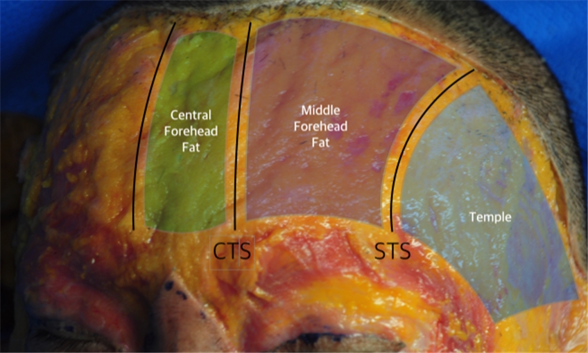
Image 3. Forehead Fat compartments(CTS: Central temporal septum, STS : Superior Temporal Septum).
References
Spiegel JH, et al. Frontalis midline dehiscence: An anatomical study and discussion of clinical relevance. J Plast Reconstr Aesthete Surg. 62: 950, 2009.
Costin BR, et al. Anatomy and Histology of the Frontalis Muscle. Ophthal Plast Reconstr Surg. Epub ahead of print, 2014.
Pessa JE, Rohrich RJ. Clinical Anatomy of the Face, Quality Medical Publishing, 2012. P. 24.
Ugur MB, et al. A reliable surface landmark for localizing supratrochlear artery: Medial canthus. Otolaryngology-Head and Neck Surg. 138: 162, 2008.
Reece EM, Schaverien M, Rohrich RJ. The paramedian forehead flap: A dynamic anatomical vascular study verifying safety and clinical implications. Plast. Reconstr. Surg.121: 1956, 2008.
Rohrich RJ. The fat compartments of the face: Anatomy and clinical Implications for cosmetic surgery. Plast. Reconstr. Surg.119: 2219, 2007.
-To be continued-


