


Superficial Temporal Artery(Frontal branch)
The STA progresses upward and bifurcates into the anterior frontal branch and posterior parietal branch. As shown in <Figure 2>, the STA sprouts from the horizontal line of the superior orbital rim in most cases. The bifurcation is above the superior orbital rim in 64% and below the superior orbital rim in 36%. The frontal branch of the STA has superomedial progression at a 60.8° angle toward the lateral margin of the frontalis muscle. The STA is still within the third layer at this point but rises superficially toward the skin surface at the superolateral quadrant of the intersection between the eyebrow upper margin and lateral canthus line(Figure 2). That is, small STA branches may exist in the subcutaneous level, medial to this area.
Facial nerve(Temporal branch or frontal branch)
To locate the temporal branch of the facial nerve, the 2D course and depth of the nerve along skin landmarks needs to be understood. Two methods are largely used to predict the course of the facial nerve’s temporal branch from the skin surface. First method is to use the frontal branch of STA described above and second is to use Pitanguy’s line. Many scholars have shown that the temporal branch of the facial nerve lies inferomedially to the STA’s frontal branch. However, a low bifurcation of the STA causes a unique variation where a few distal strands of the temporal branch rise above the STA. In the second method of using Pitanguy’s line, the facial nerve’s temporal branch passes along the imaginary line connecting the points 0.5cm inferior to tragus and 1.5cm superior to the eyebrow lateral margin(Figure 2).

[Advertisement] Ultra Skin/Pastelle – Manufacturer: WONTECH(www.wtlaser.com)
Next, the depth also needs to be assessed. Among the five branches of the facial nerve, the temporal or frontal branch has a unique course. The other four branches pass the parotid gland to progress under the deep facial fascia and insert into the mimetic muscle. However, the temporal branch penetrates the deep fascia(the DTF at the temple) to rise toward the surface. Agarwal et al. explained that the temporal branch travels through the fourth layer of LTC and rises superficially to the floor of the STF at 1.5~3.0cm above the zygomatic arch’s upper border and 0.9~1.4cm posterior to the lateral orbital rim (marked with a star in <Figure 2>)(Figure 2).
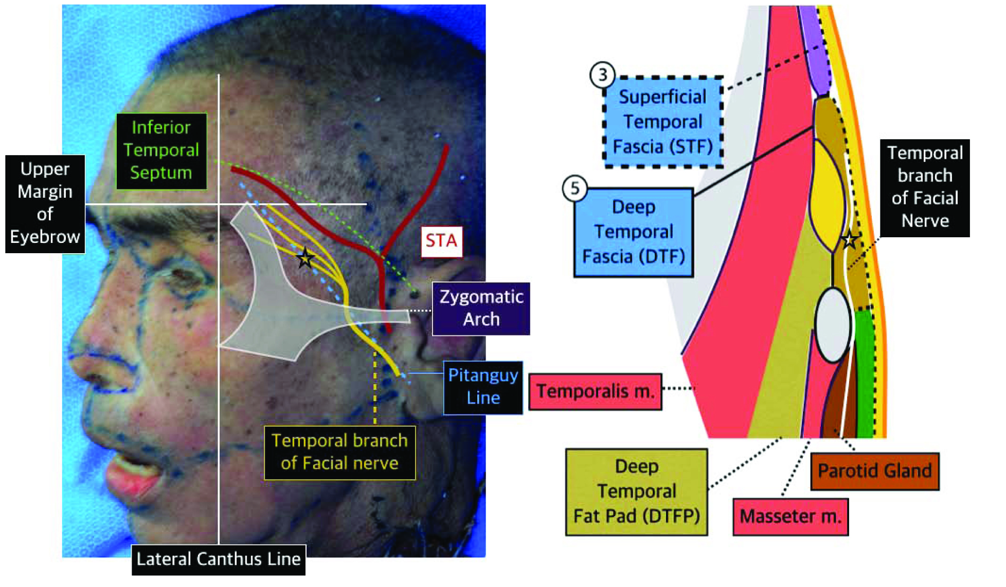
Figure 2. Position of temporal branch of facial nerve.
In the next article, we will take a look at sunken eyelids and pretarsal fullness, as the last part of our discussion on the upper face.
References
O’Brien JX, et al. New Perspectives on the Surgical Anatomy and Nomenclature of the Temporal Region: Literature Review and Dissection Study. Plast. Reconstr. Surg.131: 510, 2013.
Hwang K, et al. Attachment of the Deep Temporal Fascia to the Zygomatic Arch: An Anatomic Study, Journal of Craniofacial Surg. 10(4): 342, 1999.
Matic DB, et al. Temporal Hollowing following Coronal Incision: A Prospective, Randomized, Controlled Trial, Plast. Reconstr. Surg.121: 379e, 2008.
Agarwal CA, et al. The Course of the Frontal Branch of the Facial Nerve in Relation to Fascial Planes: An Anatomic Study. Plast. Reconstr. Surg.125: 532, 2010.
Lei T, et al. Using the Frontal Branch of the Superficial Temporal Artery as a Landmark for Locating the Course of the Temporal Branch of the Facial Nerve during Rhytidectomy: An Anatomical Study. Plast. Reconstr. Surg.116: 623, 2005.
-To be continued-


