


An aesthetically ideal eye area has appropriate height and shape of the upper eyelid crease and eyebrow with slight plumpness in the periorbital area. Volume loss or sagging around the upper eyelid can create multiple folds, giving the entire face a tired or aged look. In such cases, filler injection or autologous fat graft are carried out for volume enhancement. Right amount of pretarsal fullness are preferred by many Asian women as it creates a youthful and attractive face.
Sunken Eyelids
Sunken eyelids refer to sagging or volume loss in and around the upper eyelids. The area of the deepest indentation is called (palpebral) superior sulcus. Sunken eyelids are also known as superior sulcus deformity, sunken eye, or hollow eyelid, etc. The bone-related reasons for sunken eyelids include congenitally deep-set eyes or bony resorption. Age-related facial bone resorption occurs at differing rates depending on the area. The orbital rim tends to be absorbed more in the superomedial and inferolateral directions. For these reasons, patients without severe eyebrow ptosis have deeper hollow eyelid in the medial side compared to the lateral. However, eyebrow ptosis is more pronounced in the lateral side and with severe lateral eyebrow hooding, the lateral hollowing of the sunken eyelid may look deeper. Volume loss in the soft tissues of the upper eyelid is also caused by excessive removal of orbital fat in the upper eyelid surgery or age-related orbital fat loss leading to concaveness of orbital septum and orbicularis oculi muscle (OOM).
[Advertisement] ULTRA THIN WALL NEEDLE – Manufacturer: AESPIO(www.aespio.com)
As shown in <Image 1>, the upper eyelid has fat layers each separating the skin, OOM and orbital septum. The superior orbital fat is so thin that is not readily visible. In many cases, the OOM is immediately exposured after skin incision. A thin layer of fat exists between the OOM and orbital septum and this layer of fat is called pretarsal fat in front of the tarsus, submuscular fat between tarsus and superior orbital rim, or retro-orbicularis oculi fat (ROOF) above the superior orbital rim (Image 1). Along the deeper orbital septum, thick orbital fat surrounds the eyeball and levator palpebral muscle, etc. to aid sliding and act as a buffer.
Filler injection and autologous fat graft are the most common methods of volume enhancement in sunken eyelids. Injecting fat or filler into the superior orbital fat, the outermost layer, is not advised as this could create visible bumps on the surface. Injecting dermal filler in the orbital fat through the orbital septum has high risk of bleeding and may disrupt gliding. The most ideal injection site for filler or fat is the preseptal fat shown in <Image 1>. However, this layer is thin and the filler or fat may be injected into the OOM even if the preseptal fat is targeted. The best way to avoid this is to inject the filler or fat when you feel the resistance of orbital septum at the needle or cannula tip. This is a safe method to avoid bumps. In addition, filler or fat should be injected adjacent to the supraorbital rim (Image2).
The first reason for this is that there is a sharp bend toward supraorbital rim above the superior sulcus and injecting only a small amount of filler or fat can bring excellent effect. The second reason is that injecting filler or fat below superior sulcus to the eyelid can cause visible bumps as this area has less fat and the firm support of the tarsus pushes the injected material anteriorly. Moreover, this area folds inward during eye blinking and the patient may complain of discomfort. For these reasons, areas marked in green in <Image 2> are the ideal injection sites for fillers and fat.
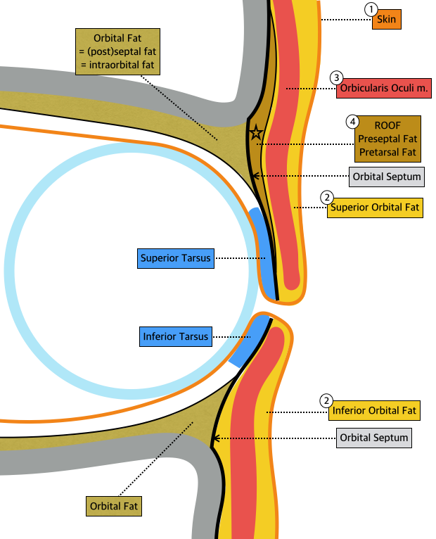
Image 1. Anatomy of upper and lower eyelids.
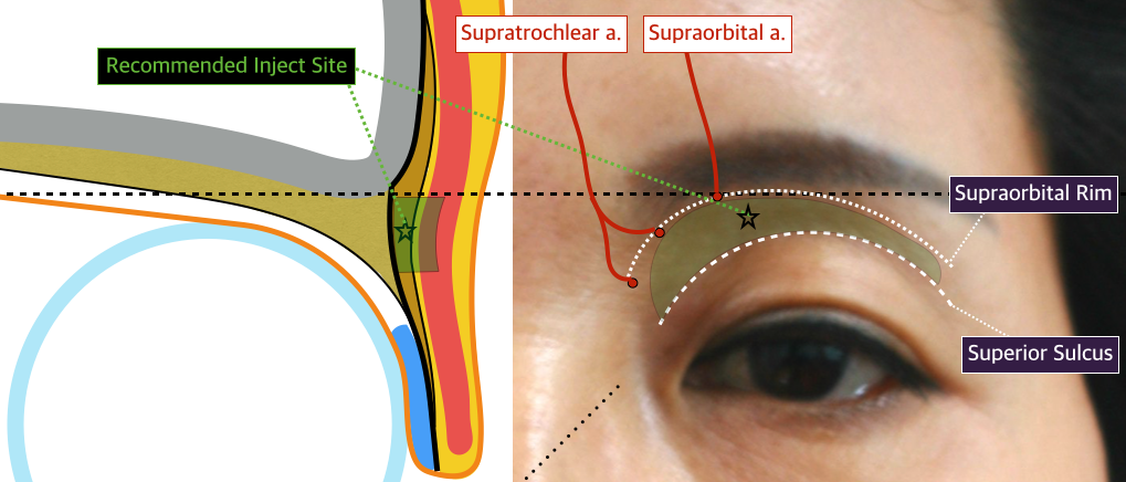
Image 2. Recommended injection site for sunken eyelid.
-To be continued-


