


6-1. The Effect of Laser Irradiation on Skin Tissues
Kawamura et al. reported in 1982 that the surface of a synthetic polymer could be etched by thiner than micrometers (10-6m) using a strong UV laser. What’s important about this study was that the remaining surface after the etching did not receive any thermal or mechanical damage, suggesting the only the target area could be removed selectively without inflicting any damage to other areas because UV laser can disintegrate the combination of synthetic polymer (photo-chemical effect). Such a clean and elaborate etching gained much attention in material processing and medical fields. In less than 1 year, Srinivasan et al. started investigating 193nm ArF Excimer laser for cutting and reshaping the cornea surface, from which current laser in situ keratomileusis (LASIK) became available in ophthalmology.
In 1983, Anderson et al. introduced a method for selective treatment of pigmentation lesions or vascular lesions with a laser without inflicting an injury to other normal tissues. Their theory was that the lesion could be selectively treated by combining appropriate laser wavelength, pulse duration and pulse energy, which is now known as ‘selective photothermolysis’. This theory has been the standard for developers of medical lasers over the last 25 years. As mentioned in the Chapter 5, laser can treat a lesion when the laser light is absorbed by various kinds of chromophores inside the soft tissue. What we need to know here is the type and properties of the chromophores that induces laser light inside the skin tissue.
When a chromophore absorbes laser light, it changes the light eneregy to thermal energy (photo-thermal effect) or to energy required for inducing chemical reaction (photo-chemical effect). But the light cannot induce any effect if it is reflected by or penetrated through the chromophore. The maximum therapeutic effect can be obtained when the laser wavelength correctly conforms with the absorption property of the chromophore. There are 3 types of chromophores in the skin, and most of the laser effects are thermal reaction, or photo-thermal effect.
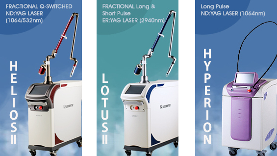
[Advertisement] ▶ HELIOSⅡ/LOTUSⅡ/HYPERION – Manufacturer: LASEROPTEK(www.laseroptek.com)
Melanosome
Melanosome is an egg-shaped pigment granule, including melanin, inside the melanocyte, which synthesizes melanin pigments. All pigmentation lesions on the skin arise from melanosome, which is why it is important to learn more about the properties of melanosome and the laser required for damaging melanosome. In order to be able to damage melanosome selectively without causing thermal injury to the surrounding tissues, we need to know their thermal properties, especially thermal relaxation time (TRT). TRT refers to the time it takes for a heated chromophore to cool down by approximately 50% in temperature and is proportional to the size of the chromophore.
Laser irradiation time shorter than TRT means no thermal injury to the surrounding tissues, allowing selective therapy. On the other hand, if laser pulse duration is longer than TRT, the heat head will be applied to the surrounding tissues, inflicting thermal injury. Melanosomes are smaller than 10-6m in size, meaning TRT approximately 10-6 seconds. Q-switched lasers are used for cutaneous pigmentation lesions for this reason. Thermal property, absorption property and the location of chromophore are required for accurate and selective treatment of a cutaneous pigmentation. Thermal property is closely associated with laser pulse duration, absorption property with laser wavelength and the location of chromophore with laser penetration depth. For example, lasers with a range of 500nm wavelength, such as frequency doubled Q-switched Nd:YAG (532nm), copper vapor (511nm) and krypton (520-530nm) lasers, have short penetration depth, limiting the treatment to the epidermis. Frequency doubled Q-switched Nd:YAG (532nm) laser has the shortest wavelength among currently available laser therapies for pigmentation lesions and yields the best result for the treatment of epidermal pigmented lesions, such as solar lentigines and freckles.
Copper vapor (511nm) and Krypton (520-530nm) lasers, on the other hand, have pulse durations longer than TRT and cause thermal injury, which is why a device for cooling down the epidermis is required for the treatment. Q-switched ruby (694nm) and Q-switched alexandrite (755nm), which have 600-700nm wavelength, have deeper penetration depth than 500-600nm wavelength lasers, allowing the treatment of epidermal and dermal pigmentation lesions. Q-switched ruby (694nm) laser is highly likely to cause post-inflammatory hyperpigmentation (PIH) after the treatment because it can strongly absorb in the melanin. 1064nm Q-switched Nd:YAG laser has the lowest absorption rate in the melanin but can penetrate the deepest (5-7mm), making it the best option for the treatment of pigmentation lesions in the dermis (Nevus of Ota, Melasma, etc.).
Hemoglobin
Hemoglobin corresponds to the chromophore for vascular lesions. The absorption peak of hemoglobin is 418,542 and 577-595nm. As mentioned above, the laser pulse duration should be shorter than the vascular TRT to not cause any thermal injury to the tissues other than the blood vessel. Vacular TRT is proportional to the diameter of the blood vessel, approximately 1-10ms when the diameter is 10-100um. Therefore, the pulse duration should be as short as 0.4-1.5ms when treating a small (~10um) blood vessel. Because the purpose of the treatment is to coagulate or shrink the size of the blood vessel, lasers for vascular lesions require high pulse energy and long pulse duration compared to the lasers for pigmentation lesions.
More specifically, the laser pulse duration and pulse energy should be changeable depending on the diameter of the vascular lesion. High energy pulse may induce rapid increase of the temperature on the epidermis while treating vascular lesion and the epidermis should be cooled down sufficiently to protect it from thermal injury. Long-pulse lasers with variable pulse duration are used for vascular lesions for this reason. Among the lasers for vascular lesions are pulsed dye laser (585-595nm), long pulse frequency doubled Nd:YAG laser (KTP, 532nm) and long pulse Nd:YAG (1064nm) laser. Dye laser can absorb a lot into hemoglobin but the penetration depth is not very deep (<1mm), which is why it is mostly used for epidermal vascular lesions, such as superficial strawberry hemangiomas or port wine stains.
KTP laser is used for various kinds of vascular lesions, including facial telangiectases and rosacea, for the largest absorption rate. Long pulse Nd:YAG laser, with less absorption rate than other lasers but almost no absorption into melanin and deep penetration depth, is appropriate for deep (~3mm depth) vascular lesions as well as small leg veins.
Water
Water corresponds to chromophore when it comes to the incision or ablation of soft tissue. The skin tissue absorbing pulsed mid-infrared laser or UV laser is primarily composed of water and collagen. The absorption peak of water is at 2.94um (Er:YAG) and 10.6um (CO2 laser), with the maximum absorption wavelength at 2.94um. The theory for incision or ablation of soft tissue is not selective treatment as in vascular lesions or pigmentation lesions; laser light is strongly absorbed in the water, causing thermal expansion on the surface of the soft tissue, and thus the ablation of the soft tissue. The laser pulse duration should be shorter than the soft tissue TRT so as not to cause thermal injury to other tissues.
Soft tissue TRT is different from epidermal and dermal layers, at about 600-800us. Er:YAG laser (2.94um) almost does not cause any thermal damage to tissues other than those irradiated at the depth of several um, because the absorption rate into the water is the greatest. Er:YAG laser can be used, therefore, for skin resurfacing without any injury to the surrounding tissues, making it the best option for the treatment of epidermal surface scar. CO2 laser (10.6um) has greater thermal property and approximately 15 times deeper penetration depth than Er:YAG laser, making it appropriate for soft tissue incision with minimal hemorrhage (thermal coagulation). However, CO2 laser may inflict thermal injury to the surrounding tissues, causing side effects such as pigmentation. When CO2 laser is used for skin resurfacing, it should have pulse duration shorter than the soft tissue TRT so as to reduce the side effects such as pigmentation.
- To be continued -
▶ Previous Artlcle : #3. Lasers and Soft Tissue Interaction Ⅰ
▶ Next Artlcle : #5. Types and Characteristics of Lasers for Skin Treatment


