Follicle stem cells in the bulge have a few important stem cell functions. First, stem cells in the bulge and upper follicle showed stronger growth capability compared to other follicle cells. Second, normal bulge cells quickly produced fast-cycling TA cells in theanagen phaseor after retinoic acid stimulation. Third, bulge cells remained undifferentiated microstructurally. Fourth, local injection of tumorigenic agent in the mouse dorsum at the beginning ofanagen phase led to increased tumor generation compared to injection in the catagen or telogen phases.
Stem cells are known to be involved in skin tumor generation. Temporary increase in proliferation of bulge cells support the hypothesis that bulge cells are stem cells and also shows that hair follicle stem cells can develop skin cancer. This is also evidence that follicle stem cells exist in the bulge area.
The bulge contains label-retaining cells(LRCs) in both humans and mice. LRCs marks DNA labels. 5-Bromo-2-deoxy-uridine(BrdU) is one of them. These cells have slow cycles, which is a key characteristic of stem cell behavior in the niche. Epidermal follicle stem cells in human hair bulge can be marked with CD200•K15 or PHLDA1. The bulge can be easily found in mouse follicle and human fetal follicles and is shown as an ORS protrusion under the isthmus. It is more difficult to find in adult follicles. In research on adult human follicles, the bulge signifies the niche for epidermal hair follicle stem cells. This is why it is difficult to clearly define the exact scope of the bulge in human follicles. The bulge is used to refer to the entire area under the isthmus and ORS. More strictly speaking, however, the bulge refers to the ORS area under the sebaceous gland in the lower isthmus adjacent to the APM.
As in the mice, it is very difficult to distinguish the stem cells in the slow cycle in immature follicle keratinocytes from progeny cells that have not started differentiating in humans. In a mouse model, it is possible to distinguish parent stem cells and progeny through lineage analysis. However, in humans, this is not possible due to ethical reasons. Currently, this distinction is dependent on traditional protein and RNA expression technology. Such methods are able to reveal a limited number of markers.
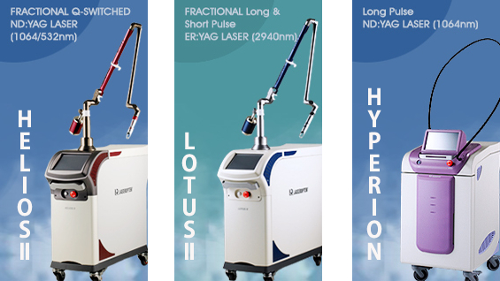
HELIOSⅡ/LOTUSⅡ/HYPERION – Manufacturer: LASEROPTEK(www.laseroptek.com)
1) K15: The biological functions of epidermal precursor cells related keratin are not yet clearly understood. K15 currently the most frequently used marker in human hair follicle stem cell research. In mice, K15 contains a label and has the capability to generate follicle cells in all phases of the cycle. In particular, at the G0/G1 stage, it has clonogenic ability. Moreover, it is also involved inepidermis and sebaceous gland generation. In mice, K15bulge cells are regarded as epidermal follicle cells.
2) K19: In humans, K19 is found in the bulge and ORS above the upper bulge and in between during follicle generation. It is also found in the basal layer of the epidermis. K19 positive bulge cells are assumed to arise from K15 cells because K15 positive cells are found among K15 and K19 dual positive cells as well as among K19 only positive cells.
3) CD200: CD200 is useful in detecting hair follicle stem cells that are expected to exist in the ORS of the human follicle bulge. In the basal layer of bulge, epidermal cells express both K15 and CD200 simultaneously but express only CD200 in the basal layer. CD200works as an immunosuppressant. It has been used in immunosuppression in both humans and mice.
4) CD34: In mice, CD34 is expressed along with K15 and is a marker for epidermal stem cells. In humans, it is not expressed in CD200+, K15+ areas. In humans, CD34 is expressed in the outermost layer of ORS under anagen phase follicle isthmus. Immunochemistry differs depending on follicle cycle and it is not expressed in telogen phase follicles. Therefore, it is noteworthy that CD34 is expressed in different cells and can vary depending on cell maturity and functional cavity. Moreover, human CD34+ cells have lower clonogenicity compared to normal hair follicle stem cells and may play a role in maturing and limiting differentiation of ORS.
5) PHLDA1: PHLDA1 is also known as TDAG51and is a proline- and glutamine-rich protein. It was first discovered in human follicle bulge in DNA microarray analysis. PHLDA1 mediates apoptosis resistance but there is controversy to this hypothesis. PHLDA1 lies in the bulge and has IR pattern similar to K15 and CD200. However, PHLDA1 is not specific to hairfollicle stem cells.
6)EpCAM/Ber-EP4: EpCAM was first discovered through Ber-EP4 antibody and is considered the only reliable marker for humantelogen phase secondary hair germ(SHG). SHG is K15, K19, CD34 negative but partially CD200 positive. SHG is involved in de novo generation of hair matrix in anagen phase and therefore, is thought to contain very important epidermal precursor cells. Moreover, EpCAM/Ber-EP4 is expressed under the epidermis in late catagen phase but is not observed during late anagen phase. Therefore, it can be used to locate epidermal precursor cells in certain phases.
In conclusion, knowledge on human hair follicle stem cells can contribute to treatment of alopecia as well as wound healing, regenerative medicine, and epidermal cancer treatment and prevention. Hair follicle stem cells in the bulge are expected to be used in treatments related to follicle synthesizing drugs or nano particle gene transfer.
-To be continued
References
1.Purba TS, Haslam IS, Poblet E, Jiménez F, Gandarillas A, Izeta A, Paus R.Human epithelial hair follicle stem cells and their progeny: current state of knowledge, the widening gap in translationalresearch and future challenges.Bioessays. 2014 May;36(5):513-25.
2. Yang CC, Cotsarelis G. Review of hair follicle dermal cells. J Dermatol Sci. 2010 Jan;57(1):2-11.
3. Ohyama M, Hair follicle bulge: a fascinating reservoir of epithelial stem cells. J Dermatol Sci. 2007 May;46(2):81-9. Epub 2007 Jan 5.
4. Lavker RM, Sun TT, Oshima H, Barrandon Y, Akiyama M, Ferraris C, Chevalier G, Favier B, Jahoda CA, Dhouailly D, Panteleyev AA, Christiano AM.Hair follicle stem cells.J Investig Dermatol Symp Proc. 2003 Jun;8(1):28-38.
5. Lee JY, Kim EY, Se Park SP, A Study on Basic Research of Hair Follicle Stem Cell Journal of Investigative Cosmetology 2012 8(2);75-80.
6. Yang CC, Cotsarelis G. Review of hair follicle dermal cells. J Dermatol Sci. 2010 Jan;57(1):2-11.
7. Ohyama M, Hair follicle bulge: a fascinating reservoir of epithelial stem cells. J Dermatol Sci. 2007 May;46(2):81-9. Epub 2007 Jan 5.
8. SohnIB,LeeWS, Effects of Substance P on Expression of Various Factors to control Hair Growth in Human Hair Follicle Culture. Korean J Dermatol. 204 Dec;42(12):1543-1551


