Dr. Jung-whan Baek of H Plastic Surgery felt limited in his reconstructive surgery by the traditional implants not providing a perfect fit with the patient’s bone structure and causing complications. He developed ‘3D FIT Face Sculpting,’ Korea’s very first answer to this problem. 3D FIT face sculpting is an innovative reconstructive surgical technique where a customized bone structure is 3D-printed to create an implant that fits each patient’s unique bone structure precisely. Dr. Baek will share his experience and knowhow on 3D printing technology and its application in the mandibular angle, cheekbone, and cranium. We hope this series provides interesting information on the cutting-edge developments in the field of plastic surgery.
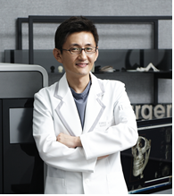
Dr. Jung-whan Baek (H Plastic Surgery)
Introduction to 3D Fit Face Sculpting
In June 2012, a female patient visited me for consultation. When I looked at her X-ray and CT images, I felt bad for her and at the same time frustrated because there was not much I could do. Some say that a doctor learns from his patients or patients are the greatest teacher to doctors but with this patient, I was at an impasse. There was little that could be done with current technology. I recommended less invasive corrective procedures and told her that I would research better options and get back to her.
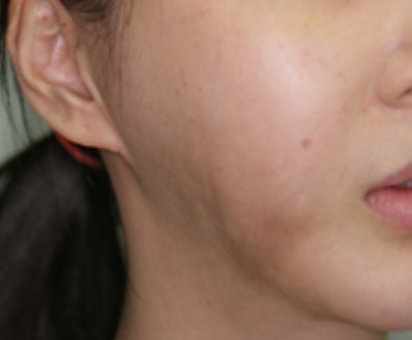

Image 1. A case of chin and jaw deformity due to drastic bimaxillary reduction surgery.
The patient suffered from jaw deformity from excessive bimaxillary reduction. It was the result of the patient’s unrealistic expectations from the surgery. She had also received a chin implant and the implant now protruded outward in what appeared as a severe underbite. Moreover, repeated silicone implant augmentation and removal as well as dermal filler injection and dissolution with hyaluronidase have thrown the facial soft tissues out of balance causing lumps and sagging. I searched for ways to reconstruct the resected maxilla but the rib transplant discussed in the textbooks was not a practical option.
A few months later, I took an X-ray of a female patient scheduled for a surgery. She was my colleague running a plastic surgery hospital in China. I was shocked to see her X-ray images. The chin implant was migrated off center and was excessively large. The angle it was inserted from was too slanted. The border of the implant could be felt during palpation of the lower face. Its distorted shape was visible at close observation.

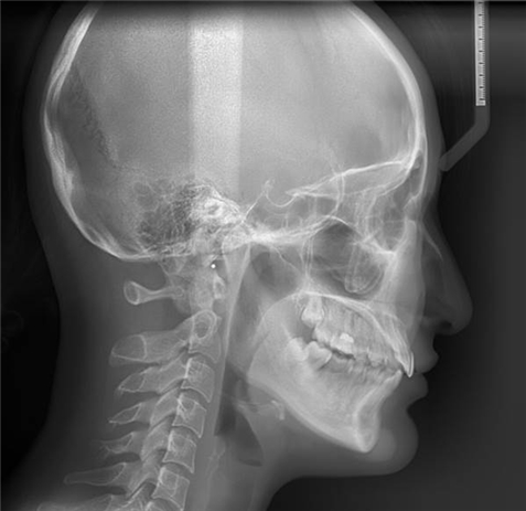
Image 2. X-ray and 3D CT images of the Chinese plastic surgeon.
The implant appeared to be in place from the side but the 3D CT image showed that it had to be corrected.
Therefore, I recommended implant removal and corrective surgery and proceeded with the bone anchoring technique to prevent future migration of the implant. However, due to the limitations of traditional implants, it was impossible to fabricate an implant that provided a perfect fit with the patient’s bone structure. I started to wonder about ways to create a customized bone implant. I believe many surgeons performing facial implant procedures shared this frustration.
Another few months passed and I encountered another patient whose X-ray reminded me of the limitations of the traditional facial contouring surgery. The question I had at the time was if a satisfying outcome could be maintained 3-4 years after V-line T-osteotomy. Even if external appearance may be normal, the bone resorption that can be felt at the finger tip along the surgical site was a serious iatrogenic complication that gave psychological distress to the patient. The jagged outline of the jaw or holes appearing from bone resorption could be somewhat filled by fibrocytes but other more serious complications could not be sufficiently resolved with currently available techniques. Was this the inevitable price the patients had to pay for a narrower jawline?


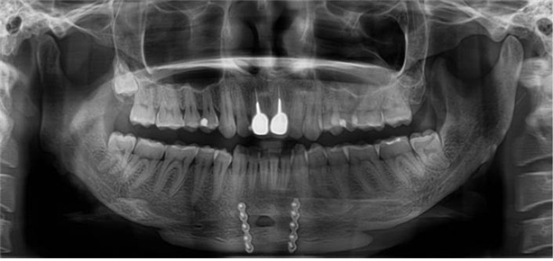
Image 3. After T-osteotomy for V-line jawline.
[Advertisement] MAGNUM(Q-switched Nd:YAG Laser) – Manufacturer: (www.i-dana.com)]
-To be continued-


