


Dr. Jung-whan Baek (H Plastic Surgery)
.jpg)
Image 9. Design of final implant II.
The area marked with an arrow in <Image 9> shows the contact area of the implant and maxillary sinus mucosa. This contact area should be solid without pores to limit the frequent inflammation of maxillary sinus from spreading to the rest of the face.
.jpg)
Image 10. 3D printed final implant.
After performing a final examination of the site and angle of implant insertion and fit using the 3D printed implant and bone model, the surgery was carried out (Image 10). The errors inherent in 3D CT imaging, image processing, and 3D printing should be considered. The model and printed implant may be deemed suitable based on pre-surgical calculations but may fail to be inserted due to hard scar tissues and other soft tissues that are not considered during 3D CT imaging. We tried to incorporate all variables that could occur during surgery and establish ways to tackle them in the planning phase.
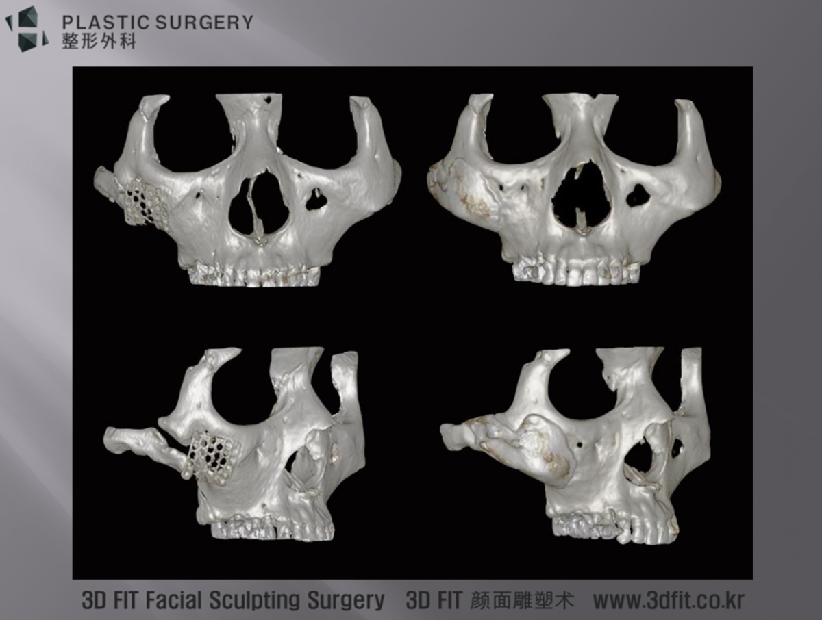
Image 11. 3D CT image before and after surgery.
The frontal and right side view show that the defect is filled without any gaps (Image 11). The boundary seen in 3D CT image is less than 1mm and the error range was less than 1mm. However, there was a slight discrepancy between the 3D CT images and the actual titanium implant’s fit in the area of defect during surgery. The implant had to be revised during surgery. To remove the protruding bone in the arch, an incision was made in the sideburn region for guiding and the implant was inserted into the desired position intraorally. The implant was firmly fixed onto the bone using three point screws.
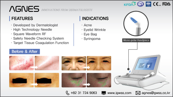
[Advertisement] AGNES(Radio Frequency) – Manufacturer: (www.igwss.com)
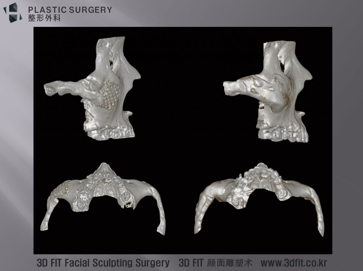
Image 12. 3D CT image.
The arch and lateral side have been reconstructed according to the initial plan (Image 12). The artifact from metallic fixtures used around the arch caused some noise but good symmetry was achieved in the outcome. As this was the first case of zygoma reconstruction using 3D implant, we went through many trials and errors. As the zygoma area had free curve and complicated defect structure compared to the cranium or mandibles, the pre-surgical preparation took a much longer time. However, the surgical outcome reflected the hard work. I believe the success of reconstruction would not have been possible without 3D printing.
-To be continued


