


Dr. Jung-whan Baek (H Plastic Surgery)
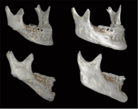
Image 4. The 3D CT image shows elongation and advancement of the right gonial angle and chin.
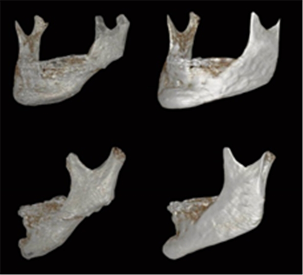
Image 5. The left gonial angle has been restored and the chin is advanced.
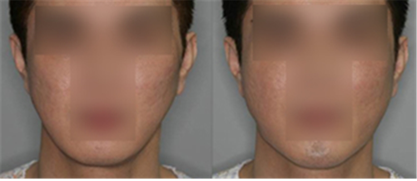
Image 6. The frontal view shows that the dents and chin asymmetry have improved.
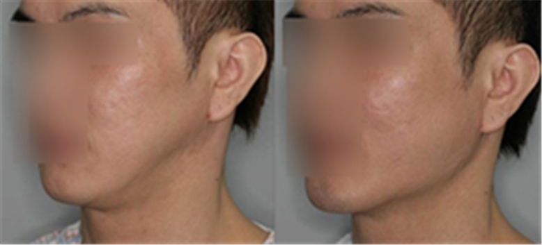
Image 7. The left diagonal view of the face shows a restored gonial angle. A slight indentation of the soft tissues remains as the masseter muscle has not extended over the enlarged bone structure. This can be corrected through soft tissue adjustment such as fat graft, etc.
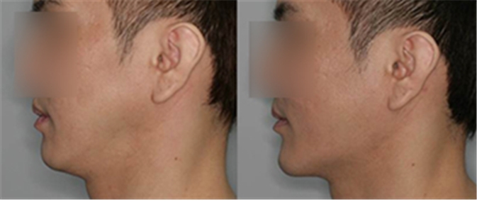
Image 8. The patient wanted a slight protrusion of the chin for a masculine appearance. The tip of the chin was advanced with the 3D Fit PMMA implant. The before and after images show the advancement. The advancement also seems to have improved the cervicomental angle.
The most important aspect of gonial angle reconstruction is the accuracy of the CT image.
The general ConeBeam CT tends to have more noise on the right side than left and is prone to distortions. Metal implants can cause large errors. Therefore, software control is needed to minimize artifacts and obtain the most accurate 3D image. The precision of the image is directly related to the surgical precision.
One aspires to 100% precision but even 3D printing technology cannot rule out all errors. The externally visible change is the result of both the change in the bone and soft tissues. Adjustment of the bone structure alone cannot address the soft tissues. As change in soft tissues cannot be predicted or measured, even surgery based on precise measurements and modeling cannot always bring the exact predicted outcome.
Virtual plastic surgery was introduced 10 years ago to predict the surgical outcome. However, these software programs are not currently used due to poor precision. Virtual plastic surgery images may even mislead the patient and instill false expectations.
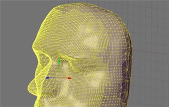
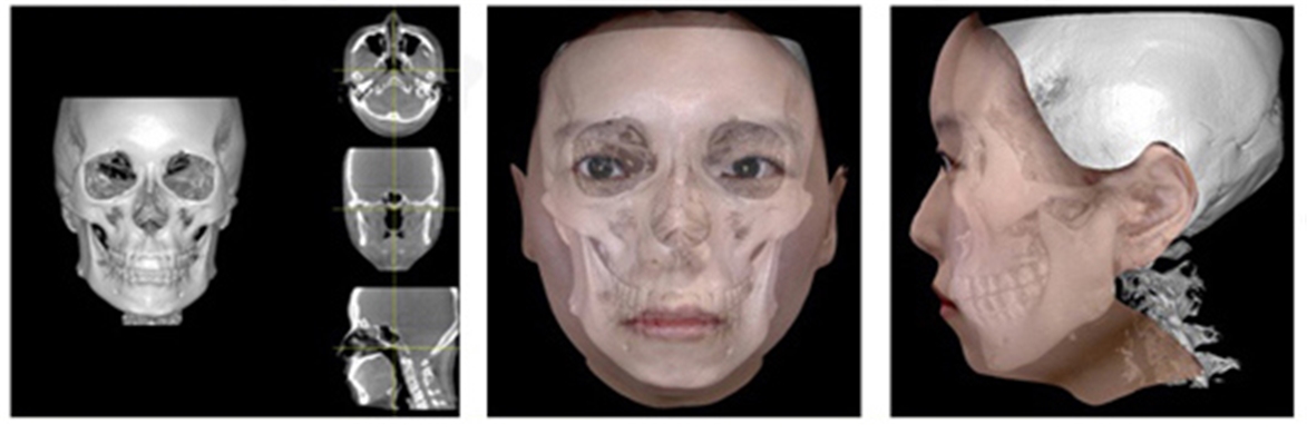
Image 9. 3D scanner is used to create a 3D map of the facial terrain. CT and 3D scanner are used to obtain data on changes of the bone and soft tissues.
Thanks to the advent of the high-precision 3D scanner, accurate 3D mapping of the facial terrain has become easy. Objective data can be used to analyze external changes. In other words, we can obtain precise data on bone and soft tissue changes and use this as a scientific foundation to analyze the interaction of the bone and soft tissues.
However, vast amounts of data need to be processed and there is incomplete understanding of the mechanical properties of soft tissues such as muscle, fat and skin. Therefore, it is still impossible to accurately predict the external outcome before the surgery using this method.
Prediction of the outcome still relies mainly on the doctor’s experience. I hope deep learning technology based on big data can enable more accurate prediction of the surgical outcome in the near future. We will take a look at deep learning in the next series of articles.
[Advertisement] ULTRA THIN WALL NEEDLE – Manufacturer: AESPIO(www.aespio.com)
-To be continued-


