


Dr. Jung-whan Baek (H Plastic Surgery)
In this issue, we will take a look at the future directions the 3D printing technology.
The 3D printing technology can be applied to various medical treatments. However, it needs to overcome a few technical and regulatory hurdles to be able to widen its application further and bring better results. I suggest the following potential solutions (Table 1).
1. Development of biocompatible materials.
2. Development of printers capable of 3D printing new materials.
3. Proven biocompatibility and safety.
4. Legal and regulatory support.
Table 1. Problems to be overcome for 3D printing technology.
Numbers _ and _ in <Table 1> have seen drastic improvements thanks to the efforts of scientists and engineers. However, this new technology should be safe and biocompatible to be used in clinical practice. Clinicians can help establish scientific data in this regard. Research is being carried out in various countries and their results are being introduced in the media. In Korea, however, due to lack of government authorization and insurance coverage, the use of 3D printing is severely limited.
I would like to discuss a patient who visited my practice but could not receive treatment due to regulatory limitations, despite her severe condition. As seen in <Image 1-1>, the patient suffers from severe bone loss and resorption in the mandible due to failed sagittal splitting ramus osteotomy (SSRO). She presented complete loss of the right mandibular condyle. Bone defect was supplemented by two bone grafts using the ilium and rib bones. She had received a commercial titanium joint implant.
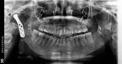
Image 1-1. This panoramic X-ray image shows implantation of various metal pieces from previous SSRO and chin osteotomy. An artificial joint has been fixed onto the right condyle with four screws. The previous surgeries were performed for the goal of aesthetic improvement but resulted in unwanted disfigurement, barely preserving the functionality. It was one of the most difficult challenges of my surgical career to tell this patient that surgery was impossible due to legal and regulatory limitations. Let’s take a closer look at the patient conditions.
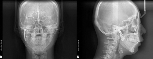
Image 1-2. The frontal X-ray image reveals artificial joint implants and longer implant placement on the left compared to right. In the lateral X-ray, the difference between the overlapped right and left mandibles was found to be over 8mm. Even in plain sight, the patient had a clearly asymmetrical and twisted lower face with drastically different condyles.
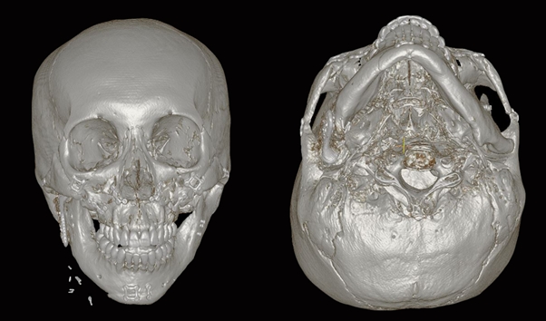
Image 1-3. The 3D CT image also shows asymmetry of the bone structure. The image on the right shows asymmetry not only of the face but the entire cranium. The central line from between the eyes, through the nose and cervical spine is tilted, showing this patient has congenital asymmetry as well.
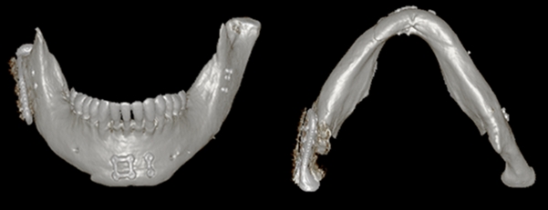
Image 1-4. Isolating the mandible shows clear asymmetry and the frontal view shows that the right condyle is markedly shorter than the left. The view from the bottom shows intact right side of the mandible whereas the left is concave.
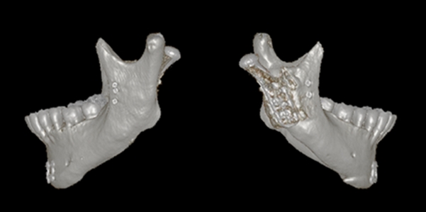
Image 1-5. From the lateral view, the artificial joint is not aligned with the normal condyle. This may be due to inadequate length and placement of the implant. Also, such a drastic osteotomy left insufficient bone structure, making the correct placement of the remaining bones difficult.
[Advertisement] ULTRA THIN WALL NEEDLE – Manufacturer: AESPIO(www.aespio.com)
-To be continued-


