None of the above hypotheses can clearly explain the formation of the tear trough. Because of the previous dissection techniques, the existence of tear trough ligament was not known till recent days. This finding explained the exact anatomical cause of the tear trough and sheds new light on considerations when designing procedures to correct the tear trough.
The Ligament
Depression below the eyes can be divided into two parts: the tear trough and the palpebro-malar groove. The tear trough is the groove extending infero-laterally from the medial canthus to the mid pupil line. The palpebro-malar groove extends from the mid pupil line to the lateral canthus. It was found that the palpebro-malar groove arises as a result of fixation provided by one of the facial retaining ligament: the orbito-malar ligament. However, the tear tough ligament was not known to the surgeons or pathologists until 2012. The reason why this important ligament was not identified is that the traditional way of dissection easily destroyed this ligament without notice. Through different approach, the tear tough ligament was finally proved to exist.
This ligament is less than 0.5 mm in thickness and 8mm (6 to 10 mm) in width, located medially between the origins of the palpebral and orbital parts of the orbicularis oculi muscle, and continues laterally from the mid pupil line as the orbicularis retaining ligament. The medial part of this ligament is 7mm (5 to 10 mm) in length, spreading from the medial canthus of the eye, and located inferior to the anterior lacrimal crest, then fixed to the dermis. The lateral part of this ligament is 16mm (14 to 18 mm) in length. This septum-like ligament was extremely strong, with histologic features of highly organized and densely packed collagen bundles, identical to the zygomatic ligament. The tear trough ligament has the effect of tethering and holding the orbicularis oculi and malar fat pads down onto the skeleton of maxilla.
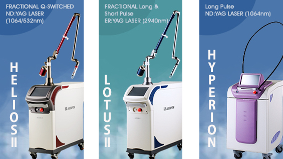
HELIOSⅡ/LOTUSⅡ/HYPERION – Manufacturer: LASEROPTEK(www.laseroptek.com)
The Treatment Procedure
Traditionally, the correction of tear trough is trough non-surgical techniques, which are mostly volume based, including the fat transplantation or hyaluronic acid injection. However, these injections often caused some problems, like Tyndall effect or unevenness of the surface. It is because the injection level should not be placed above or directly into the tear trough, as this can aggravate the tear trough. The fillers should be placed in the area right below the tear trough ligament. In general, these techniques are only effective for patients with mild tear trough presentation and slight protrusions of the lower eyelid fat pads. The effectiveness of fillers in softening the tear trough is due to restoration of the volume loss underneath this ligament.
In order to achieve better results of tear trough correction, we should at least try to ameliorate the tethering effect of the ligament other than injecting fillers only. If we can first release the tear trough ligament completely or partially by using blunt cannula or subcision, the elevation of the mid cheek as well as correction of tear trough can be achieved at the same time. Subsequent injection of hyaluronic acid or fat will result in more volume restoration and prevent reattachment of the tear trough ligament to the maxilla. This minimal invasive procedure of ligament adjustment with filler injection was proposed initially by some dermatologist (Dr. Shiou-Han Wang) in Taiwan and now is gaining its popularity here through recent years.
.jpg)
Figure 1: Before and after tear trough ligament adjustment with filler injection. (Courtesy of Dr. Shiou-Han Wang)
This technique improves the tear trough by using an 18 gauge 70mm blunt cannula inserting from the lateral cheek directly to the point below the tear trough and fanning technique firstly to release the tear trough ligament along with linear threading injections of hyaluronic acid right beneath the ligament. The physicians should avoid injections into the orbicularis oculi muscle accidentally to avoid lumps or bumps when the muscle contracts.
.jpg)
Figure 2: Before and after tear trough ligament adjustment with filler injection. Note the entry point of 18 gauge needle at left cheek. (Courtesy of Dr. Shiou-Han Wang)
The advantage of this procedure is immediate correction of tear trough without Tyndall effect, surface irregularity, or bruising (Figure 1 and 2, courtesy of Dr. Shiou-Han Wang). However, the duration of this ligament adjustment with filler injection is similar to the traditional filler injection only, which depends on the properties of various fillers (such as hyaluronic acid, collagen, or fat). It is recommended that a more long-lasting gel-type hyaluronic acid with a higher degree of cross-linking (such as Juvederm Volbella or Voluma) would be the better choice for injections right beneath this released ligament.
-To be continued


