Dermatologic surgery complications include bleeding, hematoma, infection, necrosis, nerve injury, suture associated complications (ruptured sutures, foreign body reaction, suppuration, etc.), dehiscence of operation scar, hyperpigmentation, hypertrophic scar, and free-margin distortion (around the eye, nostril, earlobe, etc.). Among these, the most common and serious complications are bleeding, infection, necrosis, and scar. Many factors are involved in the cause of these complications with the operating doctor’s medical knowledge (knowledge on anatomy and complications) and surgical techniques (experience and skills) being the most common cause. In order to be able to prevent complications and to take appropriate measures immediately once complications occur, it is imperative to first know your own limits. Efforts to continuously train are important. Learn the surgical techniques and experiences discussed in textbooks and journals, etc. and hone your skills through applying them in real clinical cases. When you continuously complement your own skills that are earned from repeating similar procedures in clinical practice with efforts to self-educate, you will see that the incidence of even mild complications gradually and drastically decline as time passes. Below, I have summarized the major complications that may arise with dermatologic surgery, their causes and solutions.
[Advertisement] MAGNUM(Q-switched Nd:YAG Laser) – Manufacturer: (www.i-dana.com)]
Bleeding and hematoma
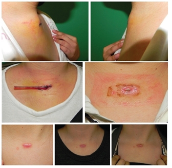
Bleeding generally progresses through the following steps; (a) early phase – gel-like mass, (b) 2-5 days – rubber-like coagulation, (c) 7-14 days – liquefaction, and (d) >14 days – absorption. In small hematomas, the surgical site swells up and then subsides (1-3 days) or resolves into the suture site. Large hematomas are a surgical emergency and occurs within 24 hours (usually 6 hours) after surgery. They are characterized by sudden pain and swelling. Complications include major structure damage, necrosis due to high tension on the wound site, wound dehiscence, infection, scarring, calcification and tumor, etc. In such cases, immediately resolve the tension on the wound site and control bleeding. Then, re-suture, open the wound or drain before administering antibiotics. In delayed hematoma, if it is accompanied by infection, necrosis, and wound dehiscence, open the wound to remove hematoma and perform secondary wound care prior to reconstructive procedure of the scar (Image 1). Delayed hematoma uncomplicated by infection, necrosis, or wound dehiscence can be aspirated by a 18G needle in 7-14 days (at liquefied state) and is treated with antibiotics.
Cause
Insufficient knowledge of major vascular courses (vein and artery).
Failure to examine and confirm pre-surgical patient conditions (bleeding tendency, medication, etc.)
Failure to pinpoint the site of bleeding during surgery due to inadequate use of suction devices, etc. and unnecessary hemostasis performed in surrounding areas.
Failure to close a severed blood vessel over 1mm thick with suture thread.
Hemostasis carried out without visual confirmation of the bleeding site.
Failure to cleanse with saline solution and recheck after initial hemostasis.
Insufficient pressure dressing after surgery.
Failure to check rebleeding within 24 hours after surgery.
Solutions
Once bleeding occurs, perform suction with a suction tube appropriately sized for the surgical site. Compressing the bleeding site with cotton swabs or folded gauze held with long forceps can help pinpoint bleeding site quickly.
Check the type of bleeding (arterial, venous, arteriovenous, and vessel size, etc.) and perform hemostasis appropriate to each type. The following hemostasis techniques can be used during surgery. For small blood vessels, electrocoagulation can be performed. For large vessels, clamping can be followed by suture or electrocoagulation (monopolar, bipolar). For persistent oozing, apply compression, elevation, epinephrine injection or gauze, cold compresses, Penrose drain (Image 1), or pressure dressing, etc. as appropriate for the situation.
Slight yet persistent bleeding sites should be compressed for a certain time (over 20 minutes). If that is not deemed sufficient, perform skin suture or bolster dressing with rolled up gauze.
For the site of continuous bleeding that resembles discharge, use anesthetics containing epinephrine or bosmine soaked gauze. Adhesives (fibrillar collagen) can also be used.
Discontinue anticoagulant or anti-platelet therapy. Related medications are as follows.
Aspirin (discontinue 7-10 days before surgery, can be resumed 1 day after surgery).
NSAIDS (depending on the drug’s half life, usually 3-4 days. Can be resumed 1 day after surgery).
Plavix (clopidogrel), Ticlid (ticlopidine): Discontinue 7-10 days before surgery. Can be resumed 1 day after surgery.
Alcohol: Avoid during surgical period.
Coumadin (Warfarin): Discontinue 3-5 days before surgery. Can be resumed 1-3 days after surgery.
Especially, Korean patients should be instructed to avoid ingesting ginger, garlic, ginko extract, ginseng, and vitamin E, etc.
If discontinuation of previous medication is difficult, minimize surgical resection and perform sufficient hemostasis and pressure dressing. Consider radiation therapy. Hospitalize the patient and switch coumadin to low dose heparin 1 week before surgery.
Remove all sutures in some cases and use simple lidocaine without epinephrine for anesthetics.
Check the wound again within 24-48 hours for rebleeding.
Hematoma exists in the liquid form within 48 hours after surgery. If hematoma is confirmed, drain as early as possible (Image 1).
When hematoma is found, about 5-10 ml can be removed using a 18G needle syringe.
If too much time has passed or coagulated mass is found, undo sutures and directly remove the hematoma mass and perform hemostasis on the bleeding site.
Hematoma increases the risk of infection and antibiotics must be used if hematoma is found.
Perform retests to check coagulopathy. Correct and consider resurgery when necessary.

Image 1. F/16 Hematoma occurring after removal of sternal notch cyst (discovered 4 days after surgery).
-To be continued-


