▶ Previous Artlcle : #5-1. Alopecia Areata
Pathophysiology
Hair follicles are known to have immune tolerance that is not attacked by immune cells.
In the case of normal hair follicles, there is no expression of MHC class I and II in the epithelium, and there are immunosuppressive cytokines such as TGF-β, IGF-1, α-MSH, etc.
Therefore, natural killer cells, CD4+ T cells, and CD8+ T cells are not observed around the lower part of the normal hair follicle.
[Advertisement] DUAL FINE BEAM – Manufacturer: SNJ(www.medicalsnj.com)]
On the contrary, alopecia areata hair follicles show that the expression of MHC class I and II is increased and immunosuppressive cytokines are reduced, indicating that changes occurred in the immune response regulation in the alopecia areata lesion.
Inflammatory cells observed in alopecia areata are mainly CD4+ and CD8+ T cells: CD8+ T cells mainly appear inside the hair follicles and hair bulb, and CD4+ T cells appear around the hair follicles.
With such evidence, the hypothesis that alopecia areata occurs due to the follicle attack of inflammatory cells as immune tolerance of hair follicles collapses is accepted.
Genetic predisposition appears as a family history in clinical practice.
According to the report on identical twins in the past, twins had similar alopecia areata onset timing or hair loss patterns, and the incidence of alopecia areata in the family was about 4 to 28%, which was higher than the incidence of alopecia areata in the entire population (0.1%).
Environmental predisposition can also affect the development of alopecia areata.
If a specific gene determines susceptibility to alopecia areata development, environmental factors are known to be able to incrementally determine the timing, pattern, degree of treatment response, and severity of hair loss.

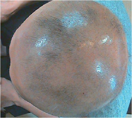
Figure 2. Alopecia totalis in a male in his 20s. Before treatment.
-To be contined


