


Histological Findings
To examine the histological changes in areas of PDO thread placement, tissue slices were harvested after sacrificing the study animals a month after thread placement. There were no noticeable differences between the controls and treated tissues in the naked eye. The navy-blue PDO threads placed in the treatment group had become clear (Figure 1).
.jpg)
Figure 1. Depigmented polydioxanone thread & dissected skin piece.
In clinical practice, in cases where suture materials have to be removed, threads placed in a patient gradually lose color and become light blue in two weeks and completely clear in 4 weeks of treatment. To the touch, these decolored threads seemed to have similar rigidity and elasticity to the thread before treatment. Histological examination of the control showed that the dermis was 1175.62μm thick and collagenous connective tissues existed in the mesh form between adipocytes in the subcutaneous fat layer, resembling the dermis (Figure 2, 3).
.jpg)
Figure 2. Dermal layer of control zone(H & E stain).
.jpg)
Figure 3. Subcutaneous fat layer of control zone(H & E stain).

[Advertisement] MAGNUM(Q-switched Nd:YAG Laser) – Manufacturer: (www.i-dana.com)]
The histological findings in the treated group is as follows.
First, during the procedure, the part of the thread inside the needle and the part outside, that is, the V shaped part, both remained in the tissue. The two hollow spaces in the cross section of the thread were visible like the eyes of an owl (Figure 4). These slides show that threads placed in the tissues do not remain in separate strands but in the folded shape, which results in two strands (one folded strand) being subcutaneously placed.
.jpg)
Figure 4. Two polydioxanone thread hollow and round granulation tissues with newly made collagen fibers(H & E stain).
Second, granulation tissues were formed around the inserted thread a month after treatment. A large amount of loose collagen fibers, eosinophil, and lymphocytes were seen in granulation tissues. Masson-Trichrome IHC (Immunohistochemical) staining, performed to observe collagen fibers, showed a large amount of newly formed collagenous connective tissues inside granulation tissues, shown as blue rings around the thread (Figure 5).
.jpg)
Figure 5. Granulation tissues and newly made collagen fibers merge with prior existed collagen fiber(Masson Trichrome stain).
Third, new collagenous connective tissues fused with the previous fibrous connective tissues to bring a merging effect. The area of suture placement went through the inflammatory response as part of the wound healing process and granulation tissues formed as a result. PMN cells, macrophages, fibroblasts, and myofibroblasts exist in the granulation tissue and release large amounts of cytokines.
Regeneration of various new tissues and cell signaling spread to surrounding tissues in the process called “mechanotransduction” and lead to the merging effect where networks are formed (Figure 6).
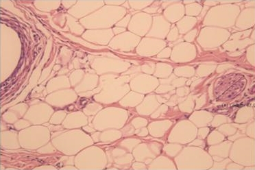
Figure 6. Mergin effect newly made collagen fiber merge with prior exstied collagen figers and connect with nerve fiber(H & E stain).
-To be continued


