


Fourth, Anti-SMA-IHC (Anti-Smooth Muscle Actin Immunohistochemical) staining showed large amounts of fibroblasts (stained blue) and myofibroblasts (stained red and in cigar-like shapes) within granulation tissues. Myofibroblasts are most deeply involved in wound contraction in the healing process and play an important role in providing elasticity and tightening effect in the treated area following RE skin regeneration therapy (Figure 7, 8).
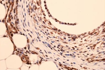
Figure 7. Blue color stained fibroblast & red color stained myofibroblast(Anti-SMA-IHC).
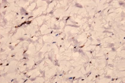
Figure 8. Red color stained myofibroblast ingranulation tissues(Anti-SMA-IHC).
Fifth, we measured the capillary area to examine an increase in blood flow. To rule out bias, we compared the largest cross-sectional capillary area in the control group to the largest capillary area of the treatment group and found that the capillary area of the treatment group (5826μm2) was over twice as large as that of the control group (2094μm2). We also observed a large amount of eosinophils (stained red) within the capillaries of the treatment group. This confirms that new collagen fibers continue to form in the wound healing process following thread placement (Figure 9, 10).
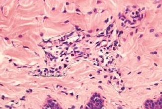
Figure 9. Microcapilary plexus(size 2094μm2) in control zone(H & E stain).
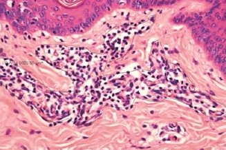
Figure 10. Microcapilary plexus(size 5826μm2) in experiment zone(H & E stain).
Sixth, porcine tissues obtained from animals sacrificed one month after treatment showed loose collagen fibers around the thread as well as adipocyte degeneration around granulation tissues. Tissues obtained from animals scarified three months after treatment showed dense collagen fibers as well as adipocyte degeneration, confirming gradual reduction of fat tissues (Figure 11).
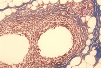
Figure 11. Denatured adipocytes around dense collagen fibers.
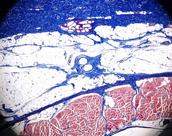
Figure 12. Fibrous bridging effect.
Seventh, dense collagen fibers formed around threads placed in the subcutaneous fat as well as new collagen fibers stretched upward to the dermis and downward to the fascia, creating a dense network resembling a spider web. This is called fibrous bridging effect, a very important evidence of tightening effect of skin regeneration procedure using PDO thread (Figure 12).
Growth Factors
Each slice of the porcine tissues obtained immediately after sacrificing the animals were refrigerated in liquid nitrogen tank. The EGF, FGF, KGF, IGF, TGF-β, and VEGF were measured in each zone of each slice. In the zones of simple sutures, we observed a mean 100-fold increase (statistically significant) increase in EGF, FGF, KGF, IGF, and TGF-β, in porcine flaps obtained a month after treatment. In tissues obtained from animals euthanized 3 months after treatment, we observed on average 10-20 fold increase in these levels. However, in the zone with 10 rotations of threads, no increase in EGF, FGF, KGF, IGF, and TGF-β was seen.

[Advertisement] Reandnè Thread Series – Manufacturer: GTG KOREA(www.gtgkorea.com)
In zones where threads were placed in the subcutaneous fat and dermis, an increased level of VEGF was seen. However, in zones of intramuscular insertion, the VEGF level increased over 10,000 times compared to that of the control. Based on such results, we inferred that simple sutures with less tissue damage resulted in longer duration of mechanotransduction. On the other hand, tissues where the threads were twisted 10 times in one direction after placement, suffered greater tissue damage and could go through short-lived mechanotransduction during the healing process.
Based on the fact we could not observe VEGF in subcutaneous fat and dermal layers of much less blood flow compared to the muscle but a greater increase in VEGF in the muscle, the release of VEGF may increase in proportion to the amount of blood flow in a given tissue. Therefore, one can consider intramuscular placement of PDO sutures in areas where VEGF action is targeted.
-To be continued


