The etiological process of vision loss could be described as follows; The internal carotid artery enters into the brain from the spinal cord and then go out from brain again. It divides into the dorsal nasal artery, supratrochlear artery, and supraorbital artery on the outside. The filler substance enters these three arteries against arterial blood pressure and obstructs the ophthalmic arterial system, which results in vision loss. Vision loss could also occur when the filler substance seeps into the damaged one of the three arteries. The angular artery that ascends from the area of the nasolabial lines joins with the dorsal nasal artery and caution should be taken to avoid damaging the angular artery.

Table 1. Frequency of vision loss per injected substance; The Korean Retina Society’s data.
Symptoms and signs
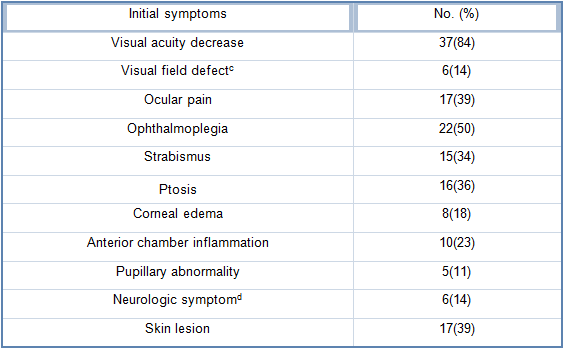
When the above conditions for vision loss are met, symptoms develop almost simultaneously with the injection in most cases. Ozturk et al. reported that even delayed symptoms will develop within 10-15 minutes of treatment. According to the Korean Retina Society’s data in 44 patients, reduced visual acuity occurred in 37 patients (84%), ophthalmoplegia in 22 (50%), ocular pain in 17 (39%), and ptosis in 16 (36%) (Table 2). Other symptoms that can develop in 1-2 days of treatment include strabismus, nausea, color change in the surrounding skin, and necrosis. Headache, pain in the ipsilateral side of the face, blurred vision, and perspiration are reported in some cases and ischemic stroke in rare cases.
[Advertisement] FCR® (Fractional Prickle CoralCalcium Regentron) – Manufacturer: (www.illglobal.com)]
Visual acuity is improved or recovered when initial symptoms are limited to partial vision loss. If complete vision loss occurs immediately after treatment, it is rarely reversible. Various remedies for vision loss are suggested such as potent steroids or hyaluronidase but most are found to be ineffective. Therefore, we should direct our efforts to prevention, not remedy.

Table 2. Rate of visual symptoms; Korean Retina Society’s data
-To be continued


