


▶ Previous Artlcle : #1-2. Pulsed Dye Laser Treatment
Classification of Vascular Malformations
Vascular malformations are categorized as Capillary (CM), Venous (VM), Lymphatic (LM), and Arteriovenous Malformation (AVM) depending on the type of affected vessels. Among these, this article will focus on the most common type, Capillary Malformation (CM).
Three out of every thousand babies are born with port-wine or dark red stains on the skin. This skin anomaly is caused by malformation of small capillaries on the surface of skin and are divided into salmon patches and port-wine stains.

[Advertisement] ULTRA THIN WALL NEEDLE – Manufacturer: AESPIO(www.aespio.com)
Salmon patches appear in the central area of the face in the form of small, pale pink lesions. They tend to spontaneously disappear after 1-2 years and do not cause a clinical problem (Figure1). On the other hand, port-wine stains, a type of capillary malformation, may continue throughout life and darken in color and enlarge in size proportional to growth of the patient. It may also progress into protruding lumps of vessels in the form of granuloma pyogenicum (Figure 2).
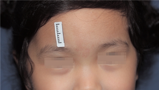
Figure 1. Disappearing salmon patches: A 2-year-old girl with light reddish patches on the forehead and between eyebrows.
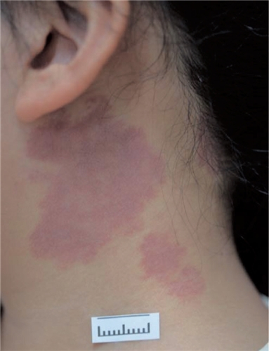
Figure 2. Port-wine stains: Reddish purple stains on the neck of a 7-year-old girl
Pathogenesis
Pathogenesis of port-wine stains has not been fully clarified. Its development is mostly sporadic to pinpoint heredity as the cause. Combined lesions of capillary-arteriovenous malformation have known hereditary cause with RASA1 gene abnormality. Therefore, the condition may be thought to somehow arise from genetic mutation. Simply put, it is considered a mosaicism caused by abnormal clone enlargement originating from the neural crest.
Clinical Presentations
In terms of circulation, port-wine stains are slow-flow malformations and as such are not higher in temperature than the surrounding skin. On the other hand, arterial blood carrying the core body heat rises to the surface in AVM lesions which are warmer in palpation.
I personally use a contact thermometer to compare the temperature between the lesion and normal skin. If there is a difference between temperatures, AVM clinically mimicking capillary malformation should be suspected. Lesions usually present as birthmarks in most cases and appear as flat patches in the early years and expand as the body surface area grows with age. These lesions progress into plaques and granuloma pyogenicum-like nodules as they thicken later in life.
Port-wine stains do not spontaneously disappear and the incidence is similar between men and women. Bleeding or ulceration are generally not observed. Most cases present a unilateral lesion but some may have multiple lesions that do not cross over the central line of the body. The coloration varies from light pink to dark purple depending on the location and depth of the lesion.
Unfortunately, these lesions appear most frequently in the face, with 40% of possibility. The facial lesions show distribution based on trigeminal nerve innervation. The lesions on the forehead and upper eyelid are affected in the area of ophthalmic branch (V1), lower eyelid, cheek and upper lip innervated in the area of maxillary branch (V2), and those on the lower lip and jaw are in the area of mandibular branch(V3).
-To be continued-
▶ Next Artlcle : #2-2. Port-wine Stain


