After such studies were published, most dermatologists hold the belief that LTID develops from impairment of melanocytes’ melanogenic function, rather than cell death of melanocytes themselves. From our clinical experiences, we discontinued LT as soon as early signs of hypopigmentation were seen and continued monitoring the patients. We found that most cases were naturally resolved. Based on our analysis of repigmentation patterns, the improvement seems to come from functional recovery of melanocytes, rather than regeneration or repopulation of melanocytes. Therefore, we agree with the melanopenic argument to a large degree.
Moreover, this argument is supported by studies showing the therapeutic mechanisms of LT are removal of melanin-melanosomes, damage to the dendrites of melanocytes, and down-regulated expression of melanogenesis-associated proteins rather than the loss of melanocytes.19,21 However, we also believe that such a claim needs more scientific evidence before it could be established as a ‘fact’.
First, the melanopenic argument is based on data from only 6 Korean patients. The data is insufficient for generalization and does not consider racial differences.
Second, the histological manifestations of hypopigmentation may vary depending on the etiology. LTID can quickly develop from an excessively high fluence that damages melanocytes or gradually develop from toxic level of total cumulative energy caused by repeated applications of a low fluence. Depending on what it is caused by, the histology of hypopigmentation can be variable.
Third, one cannot rule out the possibility that histological changes take place over time since the onset of hypopigmentation. Impaired melanocytes that are alive immediately after the treatment can later go through apoptosis, or destroyed melanocytes can be repopulated or replaced by new melanocytes.
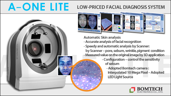
[Advertisement] A-One LITE(Facial Diagnosys System) – Manufacturer: BOMTECH(www.bomtech.net)
Although most dermatologists know that there is relatively large volume of data supporting that LTID is caused by functional impairment of melanocytes and not destruction, they should entertain the possibility of opposite views. Furthermore, they should strive to collect and analyze histological data associated with clinical information including treatment techniques, manifestations of hypopigmentation, patient conditions, and outcomes. Most cases of LTID are undesired complications of the procedures for aesthetic purposes, so patients are not likely to agree to skin biopsy. However, as non-invasive methods including reflectance confocal microscopy can be more widely used in the future, we will be able to gather and discuss much more histological data of hypopigmented tissues.
-To be continued


