Pigmented lesion over the right eyebrow
1. Case
A young female patient was 6 years old at the initial visit. She presented a pigmented lesion over the right eyebrow which she has had from birth. I tried various treatments over 2 years.
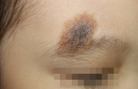
Image 1. Initial visit.
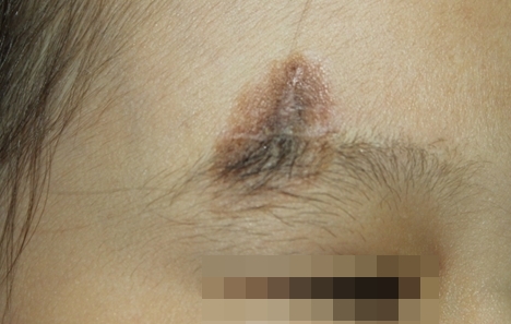
Image 2. Two months afte Er:YAG laser therapy.
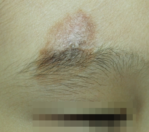
Image 3. One month after fractional CO2 laser.
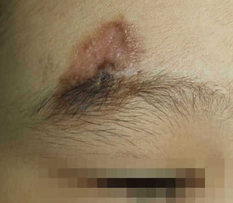
Image 4. Three months after fractional CO2 laser.
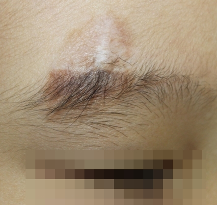
Image 5. One month after TCA 50% resurfacing+cryotherapy.
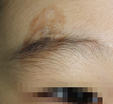
Image 6. One month after TCA 50% resurfacing+cryotherapy.
[Advertisement] ULTRA THIN WALL NEEDLE – Manufacturer: AESPIO(www.aespio.com)
2. Therapeutic considerations for the present case
1) The diagnosis is unquestionably congenital melanocytic nevus (CMN). Most patients with CMN that are around the age of 6 do not respond well to laser therapy and surgery should be considered. However, the location of the lesion in this case makes surgical removal difficult. Especially, the pigment build-up is seen across the vertical length of the eyebrow, which makes surgery more difficult.
2) The mole is thicker in the middle with dark blue coloring and spreads out into thinner brown color in the form of café-au-lait spot. As seen in the treatment outcome, and contrary to expectation, the recurrence is more frequent in lighter outer edges of the lesion.
3. Revisiting the course of the treatment
1) Was it a right decision to choose a non-surgical method?
The present case was treated with non-surgical methods including laser, cryotherapy, and chemical peel. However, the patient may have benefited more from surgical removal of the center of the lesion with thicker mole tissues as a first step of the treatment.
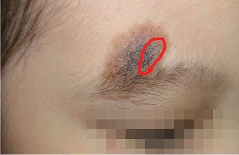
Image 7. Change into café-au-lait spot after excision
I believe that after surgical removal of the area marked in <Image 7>, the lesion would have changed into a type of brown café-au-lait spot that responds better to laser. Lim et al. reported that maximal surgical removal of the lesion followed by laser resurfacing was effective in CMN (‘A Combination of Dual-mode 2,940 nm Er:YAG Laser Ablation with Surgical Excision for Treating Medium-sized Congenital Melanocytic Nevus. Ann Dermatol 2009:21 (2)120-124]’). Likewise, in the present case, combination of surgery and laser resurfacing carried out after complete healing of sutured areas would have offered a more comfortable treatment and better outcome.
2) Is it possible to treat eyebrow lesions?
From the initial visit, I did not consider pigmented lesion inside the eyebrow a treatment target as treatment could result in partial loss of the eyebrow. However, TCA peel and cryotherapy were carried out after poor response to laser in areas including the inside of the eyebrow and led to significant improvement of the lesion. As mentioned earlier, this may be due to the fact that the part of the lesion that lies across the eyebrow is brown where melanocytes may not be lying too deep. Therefore, I believe it would have been better to try various methods that do not damage the follicles. In this case, shaving of the eyebrow under the parent’s consent would have been needed prior to treatment.
3) Was laser resurfacing the only choice available?
It is well known that the traditional laser therapy of CMN consists of delicate resurfacing using CO2 laser or Er:YAG laser. However, these methods are also known to often cause scarring or recurrence. Currently, non-resurfacing lasers that have been effectively used were Q-switched Alexandrite, and Q-switched Ruby laser, etc. The efficacy of these lasers can be limited to only CMN lesions with histologically shallow mole tissues but they offer easier post-laser care and superior aesthetic outcome compared to laser resurfacing. As the present case is a young pediatric patient and the lesion is brown with mole tissues likely lying shallow, the Q-switched laser could have been effective.
4) Combination of laser with TCA peel or cryotherapy
TCA peel and cryotherapy were useful methods that were applied before the advent of laser therapy. These two methods gave way to laser as the optimal depth of treatment was difficult to assess and this brought side effects and failure. However, when complete removal of the lesion is difficult with laser, as in the present case, combination could have brought better results.
4. Conclusion
1) In a pediatric patient, such as the present case, the treatment frequency should be as low as possible. To this end, histological examination of the center and outer edges of the lesion should be carried out to assess the depth and distribution of the lesion before treatment. This provides a more clear understanding of the lesion and helps determine the optimal treatment course. However, biopsy was avoided in this case due to the young age of the patient and this interfered with selection of the correct treatment course.
2) It seems wrong to have considered laser resurfacing as the only treatment option. In a better treatment plan, other modalities such as surgery, chemical peel and cryotherapy, etc. could have been tried first to reduce the lesion before laser resurfacing could more closely tackle the remaining lesion.
3) It was wrong to assume that lesions occurring in hairy areas such as the eyebrow should not be treated. Without treatment, be it laser or other modalities, aesthetic improvement cannot be achieved.
4) This patient did show great improvement from the original lesion but light pigment lesions continue to recur as in the treatment of café-au-lait spots. Expertise and effort are needed to treat this residual lesion.
-To be continued-


