Comments on the article ‘Dilemma’ in the last issue(#1-1. Quiz)
Clinical images from the last issue

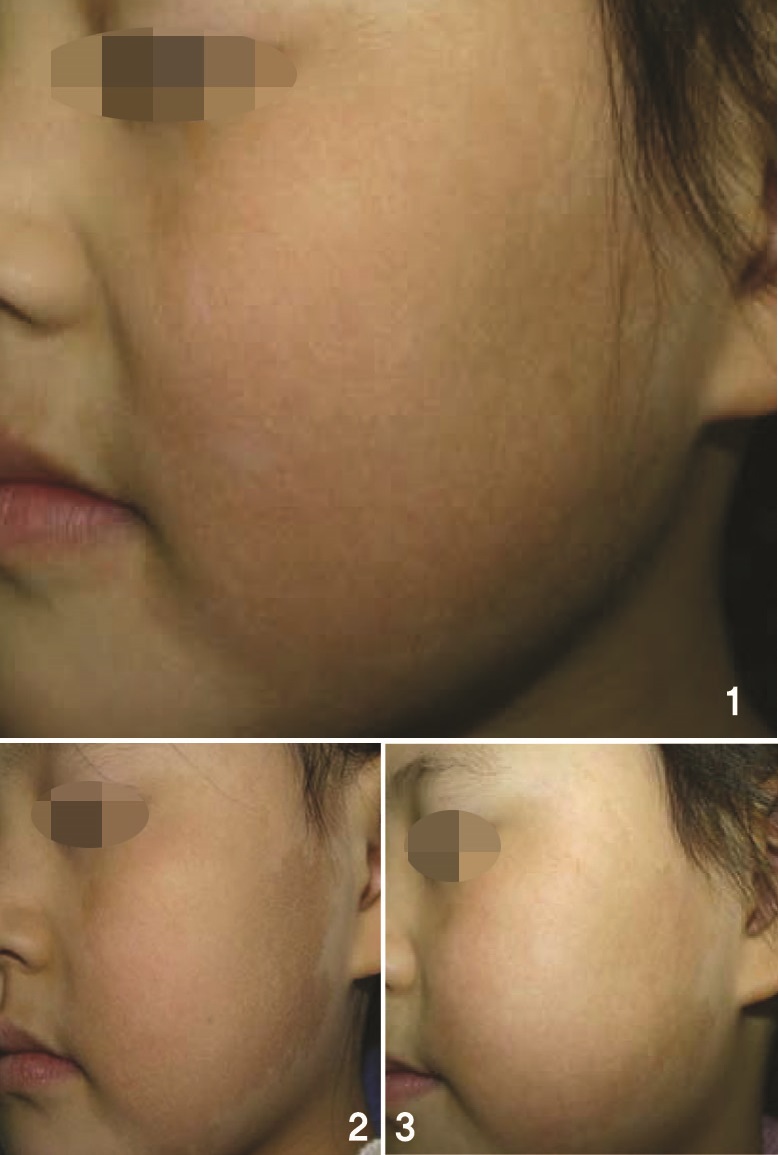
Image 1. Initial visit (2008).
Image 2. 3-5 treatments with Q-switched Nd:YAG laser (2009).
Image 3. Er:YAG, IPL treatment (2010).

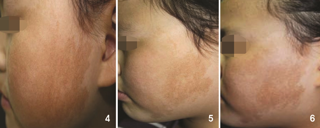
Image 4. Hair removal laser, long-pulsed KTP, etc. (2011).
Image 5. Laser toning + whitening agent (2012).
Image 6. Laser toning + whitening agent continued (now).
Take slow but steady steps forward
This is definitely one of the most difficult cases. In general, only successful cases tend to be reported in the literature (Even in these cases, the success of treatment should be corroborated through years of follow-up. Some report temporary effect as successful outcome). However, we know that there are many cases of treatment failure. Following treatment regimens introduced in the literature does not always bring the same results. This may be due to the errors made by the authors or individual differences among cases. Studies suggest one possible treatment and do not offer one-size-fit-all solution.
Clinical manifestations point toward epidermal melanocytosis. Biopsy would screen for either café-au-lait spots or Becker’s nevus. Here, the therapeutic perspective is important. For example, a paper reported that cafe-au-lait spots have a ‘dermal factor’ which stimulates epidermal melanocytes. In other words, addressing only the epidermis will not bring improvement. The dermis will also have to be tackled. This is why the treatment outcome differs from those of solar lentigines or freckles. Q-switched laser alone can bring improvement but this is rare. And it would be more unlikely in this case with faint pigmentation. Combination of Q-switched laser and CO2 laser (fractional technique) is possible but this increases the risk of scarring (it should be used as a last resort). From my experience, combination of Q-switched laser + liquid nitrogen therapy may bring partial effect but this also carries the risk of scarring and requires a lot of experience on the part of the doctor.
Regardless of the treatment modality, PIH is inevitable. The trick is not to be too aggressive and take a slow, step-by-step approach. In this case with soft pigmentation, a Q-switched laser with high melanin absorbance and short wavelength should be selected. However, one should be aware that PIH could follow over-dose or even normal dose of laser treatment. Dr. Kang Wonhyung, Q Dermatology Clinic
[Advertisement] ELLA-DUAL - Manufacturer: INSKIN(www.inskinkorea.com)
How about using a vascular laser?
Regarding diagnosis: Looking at the characteristics of the lesion in the first image (Image I) of the patient at the age of 8, the pigments definitely have thickened and as the color is close to brown rather than gray or blue, pigment increase in the epidermis or upper dermis can be suspected. Also, the strong red coloring in the center of the cheek and the change to dark red in the later image (Image 6) hint at vascular involvement. Therefore, other than the Professor Won-suk Kim’s diagnosis, I think the likely diagnosis would be, assuming that the patient received therapies including laser from another doctor prior to the first visit, pigment deposition over a mild portwine stain. Or, it could be an atypical form of erythromelanosis follicularis faciei et colli unaccompanied by follicular keratosis.
It may be necessary to identify major pathophysiological factors through histological comparison of the lesion and the surrounding normal tissues.
Regarding treatment: Becker’s nevus or café-au-lait spots are difficult to treat because they arise from congenital mosaicism affecting the mechanism of pigment formation in the lesion area rather than from actual pathophysiological causes. Therefore, pigments can be temporarily destroyed through whatever therapeutic means possible but the body reproduces pigments to return to the original state. This is why there are many cases that initially show improvement after treatment but return to the original condition. The present case may be one of such cases.
If the goal of treatment is aesthetic improvement rather than complete loss of the lesion, a vascular laser could be tried to tackle the vascular factor that causes the dark red color. – Professor Hong Seungpil, Dermatology, Dankuk University Hospital
-To be continued-


