


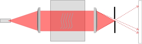
Figure 3. Diagram of ultrasound visualization.
Wontech provided an ultrasound visualization device to ensure the quality of the HIFU transducer. The three-axis control system and force balance meter are used in the final testing before the product release. The ultrasound visualization is a very useful tool for validating the transducer performance during the early phase of product development. The ultrasound visualization shown in <Figure 3> uses the German physicist Toepler’s Schlieren imaging. Toepler applied Foucault’s blade experiment to visualize the refraction of light beam traveling through a transparent medium caused by nonuniformity. The German word ‘Schlieren’ means stripes.”
The values obtained from ultrasound visualization accurately tell whether the ultrasound beam is correctly concentrated on the focal point. Moreover, imaging of the output power for analysis as well as precise measurement of thermal coagulation point’s shape and size are also possible.
The camera is installed at the location of the screen to store the images using ultrasound visualization equipment shown in <Figure 2>. Saved images go through image processing to quantify the values to be obtained. Desired images are obtained through eliminating the reference images from images measuring flow field changes. The reference image contains a standard ruler which is used to read quantified values at the focal point. The actual images taken by the camera of ultrasound visualization equipment are stored as red images produced from Laser diode as shown in <Figure 4 a>. Using color mapping, color changes corresponding output can be obtained as seen in <Figure 4 b>.
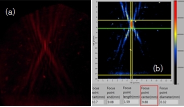
Figure 4. Image data of the transducer measured by ultrasound visualization equipment.
As discussed above, the ultrasound visualization equipment has many advantages and is essential for verifying the quality of the transducer. The opacity of the flow field which could occur from repeated use can compromise the quality of images. The opacity is caused by particles floating in the water. It is necessary to always use clean water but a large amount of images can be obtained before testing to ensure equalization and noise removal. This can be used to control the output image of the transducer.
In summary, ultrasound visualization equipment can be used to visualize the output of the HIFU transducer. This allows accurate measurement of the focal distance, size and shape of the transducer as well as the power of ultrasound output. Moreover, the three-axis control system also facilitates rapid and accurate quality testing of the cartridge of the transducer. If various measurement conditions are required, the size of the lens can be varied according to the measurement range. The output of continuous wave ultrasound can also be measured with a high-speed camera for easy adjustment.

[Advertisement] Ultra Skin/Pastelle – Manufacturer: WONTECH(www.wtlaser.com)
-To be continued-


