


▶ Previous Artlcle : #10-2. Approach to Melanonychia
Diagnosis and treatment approaches of Basal Cell Carcinoma
Diagnosis
Among different subtypes of basal cell carcinoma (BCC), nonsymmetrical, nodular, pigmented BCC with a mole-like appearance at the onset progressing into a slightly protruding, nonsymmetrical nodule form, is the most common type found in Koreans. This subtype has a concave lesion and is often accompanied with ulceration and vasodilation on the surface. Pigment appearance is not consistent, mottled with brown or black spots. Early clinical presentations of BCCare in the form of a pigmentary nodule as in a mole and only an experienced dermatologist can distinguish BCC from moles by gross examination.

[Advertisement] DEPILIGHT(808 Diode laser for Hair Removal) – Manufacturer: DANIL SMC(www.danilsmc.com)
Recently, BCC is often mistaken for a mole and treated with laser. Dermoscopy can help minimize recurrence from misdiagnosis and differentiate BCC from melanoma. Dermoscopy is more accurate in diagnosing BCC than an experienced specialist’s gross examination, however, diagnosis can only be confirmed by a biopsy. Dermascopy findings that strongly suggest BCC are known to include focal ulceration, absent pigment network, large blue-gray globules within the darkened areas, gray-blue ovoid nests, arborizing vessels, areas exhibiting spoke-wheel or maple-leaf structures, etc. Simultaneous presentation of ulceration, gray-blue globules and telangiectasia, etc. also indicate a strong clinical relevance toward BCC. Therefore, in clinical settings, dermoscopy should be performed on an early-phase lesion and if the results are found to be positive, a skin biopsy is followed to confirm diagnosis. On the other hand, a histophathological examination should be performed first on an advanced nodular lesion.
BCC cases
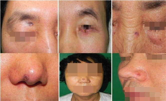
Figure 1. Images of patients diagnosed with BCC: BCC affects the T-zone of the face most frequently in Korean patients (Reference 1). In this case, a complete excision with frozen section histology should be carried out rather than a simple excision.
-To be continued-
▶ Next Artlcle : #11-2. Treatment approaches for Basal Cell Carcinoma of Face in Koreans and Mohs Micrographic Surgery


