


1. Introduction
Therapeutic ultrasound is different from ultrasound for imaging in that the latter should not inflict any change in the tissue where the energy goes through or target tissue, while the former should induce from reversible to irreversible change and denaturalization in various tissues.

[Advertisement] A-One Tab with Wi-Fi(Skin/Hair Diagnostic System) – Manufacturer: BOMTECH(www.bomtech.net)
This series will introduce the principles of therapeutic ultrasound and the way it is used for treatment through interaction between actual tissue and ultrasound energy. It has been about 100 years since the first report about the possibility of biophysical application of ultrasound. Another 50 years passed before a clinical result involving human was reported. With recent developments in image guidance systems and monitoring devices, more various therapeutic ultrasound devices are applied to clinical use. This field is still growing and a diversity of therapeutic devices are expected to be constantly developed and evolved.
In this article, we will look into the ‘Thermal and Non-cavitational Effect’ and then ‘Acoustic Cavitation’, among biophysical principles of ultrasound. These two are described separately, because non-cavitational effect induces tissue change by energy, which is directly absorbed in the tissue, generating heat or forming one-directional, radial force, while cavitation effect is associated with the activation – either imploding or non-inertial – of the generated bubble. After introducing the above principles, clinical effects based on direct interaction between ultrasound and tissue and drug and gene delivery through ultrasound will be presented.
Among the clinical applications based on direct interaction between ultrasound and tissue are ‘US mediated rehabilitation’, ‘acoustic hemostasis’ and ‘US-guided HIFU and thermal ablation’.
Ultrasound can deliver drug and gene easily by acoustic cavitation effect which enhances tissue permeability. Using US contrast agent in the process can further increase the efficiency. Sonophoresis is an application of ultrasound for facilitating percutaneous absorption of substances, such as drug or vitamin. The permeability of blood-brain barrier can be enhanced so that drug could be better delivered, or sonoporation can be used for better delivery of gene to cell. Recently, sonothrombolysis is applied to deep vein thrombosis or in combination with thrombolytic agents for cerebral thrombosis. A variety of therapeutic approaches using ultrasound is currently being investigated in these fields, some of which will be introduced briefly in this article.
2. History of Ultrasound
Thirty years after the discovery of piezoelectric effect by the Curie brothers in France in 1880, a french scientist named Paul Langevin, a student of the brothers, first observed that fish in a water tank could be killed by irradiating ultrasound. He was also the first to report pain when ultrasound was irradiated on the palm (Langevin, 1917) and first to use ultrasound to detect ultrasonic waves in water.
Langevin observed cavitation when ultrasound was discharged in water. However, sonar was first proposed by Richardson for the sinking of the Titanic in 1912, not by Langevin. In 1927, Wood and Loomis conducted more specific trials. The level of ultrasound energy could not be defined at that time, but they found that the energy level ranged from the level that could explode a paramecium to that capable of killing a small fish. In 1930, Harvey published the physical, chemical and biological activities and their involvement in denaturalization of microorganism, cell, tissue and organ.
In 1932, Freundlich et al. suggested inducing tissue heat response by using ultrasound for therapeutic purpose, and first used ultrasound for physiotherapy in 1939. Between 1940 and 1950, acoustic surgery using thermal resection by raising tissue temperature was proposed, and damaging the vestibular organ for relieving the symptoms of Meniere disease, removing breast tumor and gallstone were attempted with acoustic surgery. In the US around this time, J. J. Wild and D. Neal could obtain B-mode ultrasound image, thereby opening the era of diagnostic ultrasound.
From 1970s, objective measuring of ultrasound energy dose was of concern for therapeutic ultrasound. In 1964, Purnell et al. suggested a quantification method called ‘cataract producing unit (CPU)’ using the time it takes to develop cataract by exposing to ultrasound for a certain period of time. Johnson and Dunn in 1976 suggested quantification of energy absorbed in tissue by using mammal brain tissue in an idea called ‘energy absorbed per unit volume’. From 1980s, a lot of studies had been conducted in the US and UK for objective quantification of ultrasound energy, before polyvinylidenefluoride (PVDF) hydrophone was acknowledged as the standard measurement. This method is still recognized as the international standard quantification method.
3. Basic Concept of Ultrasound
Ultrasound is a type of sound wave and is an energy delivered by the vibration of medium molecule. Sound wave is divided to the interval where the molecules are compressed and the interval where the molecules are dispersed, and transmits energy by repeated rarefaction. Types of sound wave include longitudinal wave and transverse wave.
Longitudinal wave vibrates in the same direction of the energy progress. Ultrasound is mostly delivered in the form of longitudinal wave in human body. The transmission speed of the longitudinal wave is faster than that of the transverse wave. Transverse wave vibrates perpendicular to the direction of energy progress. An example is the ripples on the surface of the water formed when a stone is thrown in the water. Transverse wave may not be generated inside the water or air.
The basic unit of dividing sound wave is frequency. Frequency is the number of waves occurred per unit time; 1Hz means 1 wave occurred in 1 second. Higher frequency is recognized as a sharp sound, and lower frequency as a dull sound. The frequency band that is recognizable by human ear is called ‘audible sound’, which is between 20Hz and 20kHz. Sound below 20Hz is called infrasound and that above 20kHz is called ultrasound.
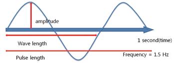
Figure 1. The standard unit of sound wave is frequency.
Medically used ultrasound devices mostly use frequencies between 1MHz and 15MHz. Therapeutic ultrasound devices particularly use frequencies around 1MHz, and diagnostic ultrasound devices between 2MHz and 15MHz. The major differences between the ultrasound imaging devices using each frequency band are resolution and penetration depth. Higher frequency means better resolution but shallow penetration depth. Wavelength (f) is inversely related to the frequency (λ). A particular medium is known to have consistent velocity of propagation (c) – for example, velocity of propagation is 1540m/s in the water – and it was revealed that c=f λ.
Attenuation is in linear relationship with frequency. Attenuation coefficient in soft tissue is 0.7dB/cm-MHz; if attenuation factor is 0.7dB/cm at 1MHz, then the attenuation factor at 2MHz is 1.4dB/cm. Higher attenuation factor naturally means shallow penetration depth.
Let’s look into several principles of ultrasound imaging devices, because these principles are also applied to therapeutic ultrasound devices as well.
First of all, axial resolution refers to the ability of differentiating two structures placed in the direction of energy progress (axial direction). Quantitatively, this refers to the minimal interval between the two distinct structures. Greater axial resolution level means decreased resolution, and lower resolution level means greater resolution capability, even of very small interval between two objects. Resolution increases with as pulse length is shortened, and is enhanced in proportion to frequency (resolution level is decreased). The longer the pulse length, it is impossible to differentiate two structures included in one pulse. Lateral resolution refers to the ability of differentiating two structures placed in perpendicular to the direction of energy progress. The beam width of focused ultrasound is lateral resolution. As the focus area has the narrowest beam width, this is where the lateral resolution is the greatest. The beam width becomes wider as the distance between the launching area of ultrasound and the focus area is longer, and becomes shorter when the frequency is higher.
The parameters and units of ultrasound is presented in <Table 1>.
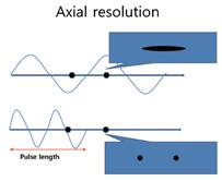
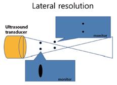
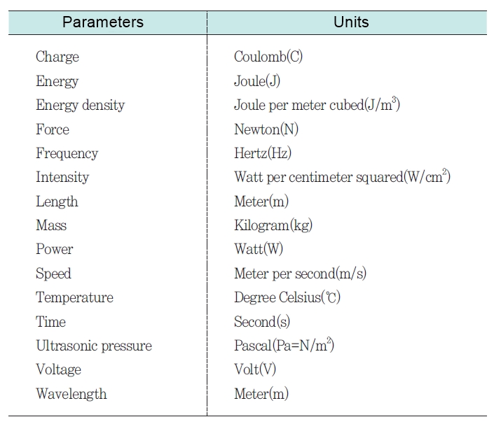
Table 1. Parameters and Units of Ultrasound.
-To be continued-
▶ Next Artlcle : #2-1. Thermal Effect and Non-Thermal, Non-Cavitational Effect of Ultrasound


