This series is intended to investigate the history, theory and indications of medical lasers from the user’s perspective. An explanation about lasers from a clinician who uses lasers in clinical settings will be a practical help to readers. The author of this series is Director of JMO Dermatology, Wooseok Koh. As an authority on hair removal treatment in Korea, Director Wooseok Koh is well known for having profound knowledge in medical lasers to such an extent as to have obtained patents in laser devices. He also participated in a collaborative research with Dr. R. Rox Anderson, a pioneer in skin laser device, while working as a fellow in The Wellman Center for Photomedicine in Harvard Medical School in the department of Dermatology. Director Koh will vividly deliver his knowledge and experiences in lasers to the readers.
When I was asked to write a series about lasers, I couldn’t help worrying where to start and what to write. As anyone would do before writing, I was not certain if I was qualified for this. But I decided to write this series in the hope that this would be a good opportunity to look back my own thoughts on cutaneous lasers and that a chronological description on the development of cutaneous laser would be of help for clinicians. Nothing happens in one day, but what happened in the past looks as if they occurred individually, in the eyes of people who did not live in that period, due to the limited memory of humans. As an old saying that ‘Rome wasn’t built in one day’, we might say that ‘cutaneous laser was not made in one day’. There might be some parts in this series where I put in a wrong date for my personal experiences in the past due to a slip of memory, but I will try my best not to make such errors affect the overall flow of this series.
[Ad. ▶ COPPER BROMID(Yellow/Green Laser) – Manufacturer: BISON(www.bisonmedical.com)
Birth of Selective Photothermolysis
Before laser (Light Amplified by Stimulated Emission of Radiation) was first realized as a ruby laser in 1960 by Maiman, it had gone through physical development not easily understandable for physicians. It is well known that Einstein played a critical role in the process.
Maiman did not develop the whole concepts of laser alone, but he is the one who practically realized the first laser. R. Rox Anderson would be the one who did a similar role for cutaneous laser. The concept of ‘Selective Photothermolysis’, also the title of an article published by R. Rox Anderson and John Parrish in the Science in 1983, was a concept developed through various stages, not an original idea that suddenly came into the world. More specifically, Dr. Anderson created the concept in 1983 by organizing existing ideas, with a little added creativity, and giving it a proper name like the icing on the cake. Since then, the concept of Selective Phototermolysis (SP) has become the foundation for the development and description of almost every cutaneous laser.
Back in the day, research papers on medical lasers can be found from 1960s, but systematic studies starts to be found from 1970, possibly because lasers were studied mainly for military purposes in 1960s. All efforts were focused on developing a laser weapon or lauching apparatus at that time but such studies were at a standstill without a tangible result. In the late 1960s, a lot of scientists turned to the medical world when it became difficult to receive any more research funds for military purposes.
For this reason, studies or textbooks on medical lasers have started to be published from 1970s, although sporadic. Leon Goldman was interested in lasers so much as to publish a textbook titled ‘Lasers in Medicine’ in 1971. He was the first executive director of American Society for Laser Medicine and Surgery and his name is on the award presented by this society. Various but sporadic studies on medical laser over the course of 10 years were focused on the skin and eyes in the early 1980, because laser pulse can be easily delivered to the skin and eyes, although it is difficult to be delivered to targets inside the body. In 1981, R. Rox Anderson and J. Parrish published two important studies on laser titled ‘The optics of human skin (J Invest Dermatol.)’ and ‘Microvasculature can be selectively damaged using dye lasers: a basic theory and experimental evidence in human skin (Lasers Surg Med)’.

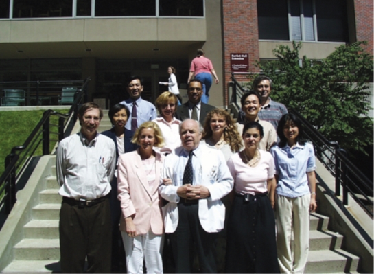
In 1999, at the entrance of The Wellman Center for Photomedicine: R. Rox Anderson (far left of the front row), Thomas B. Fitzpatrick (third from the left in the front row), and the author (right side of the third row)
At that time, Dr. Parrish was a professor of Dermatology at Harvard School of Medicine, and Dr. Anderson graduated from MIT and, after working as a science teacher at a secondary school, had been working with Dr. Parrish as a researcher for several years at the Wellman Center for Photomedicine, an affiliated organization of Harvard Medical School. Dr. Anderson entered Harvard Medical School in the mid 80s and completed his dermatology residency at Harvard. He is now Harvard Medical School Professor in dermatology, but it is interesting to think that he was not a physician or a professor but just a researcher in 1983.
The above two studies introduced two important concepts in cutaneous lasers. One of them is ‘therapeutic window’, and the other is ‘selective damage’. The former refers to the possibility of delivering light energy to even deep and widely distributed chromophore at the wavelength range that can minimize scattering by collagen, and the latter refers to the possibility of selecting a proper laser light for destructing blood vessels inside the skin while minimizing injuries to the surrounding tissues and epidermis. These two concepts become the basis of the study ‘Selective photothermolysis: precise microsurgery by selective absorption of pulsed radiation (AndersonRR, Parrish JA.)’ published 2 years later in the Science.
The study ‘Selective Photothermolysis’ provides basic concepts which are important for the understanding of cutaneous lasers; however, misunderstanding these concepts as absolute may work as a barrier in understanding clinical effects and side effects. Therefore, it would be helpful for overall understanding of this study if this term was switched to ‘relatively selective photothermolysis’ on the assumption that this is not an absolute theory to cover every case. This is an excellent study which is helpful for any clinicians utilizing lasers for skin therapy, and can be obtained easily on the internet. Below is the abstract of this study:
'Suitably brief pulses of selectively absorbed optical radiation can cause selective damage to pigmented structures, cells, and organelles in vivo. Precise aiming is unnecessary in this unique form of radiation injury because inherent optical and thermal properties provide target selectivity. A simple, predictive model is presented. Selective damage to cutaneous microvessels and to melanosomes within melanocytes is shown after 577-nanometer (3 x 10(-7) second) and 351-nanometer (2 x 10(-8) second) pulses, respectively.'
In summary, this is a simple theory that radiation of a relatively better absorbable pulse to a target within a short period of time provides selective damage to the target while preventing injuries to the surrounding tissues. The relationship among selective damage, radiation time and wavelength is well documented in this study. This theory itself is not sufficient to be applied for a procedure since the study does not highlight the fact that the condition of surrounding tissues differs between people and races and that even the same target may have different size and depth in each individual. Nevertheless, there is no question that understanding this theory well is the basis of understanding cutaneous laser. This theory triggered the development of vascular lasers from argon laser, which would leave scars a lot, to dye laser (Flashlamp Pumped Pulsed Dye Laser). This theory was also the basis applied for the development of lasers for pigmentation disorders, as well as lasers for skin resurfacing and hair removal.
Early lasers for vascular diseases have been developed to target mainly nevus flammeus in children due to the difficulty of selecting a radiation time and wavelength, because the radiation time of a dye laser is too short to damage thick vessels.
The development of lasers for vascular lesions since 1980s will be presented in the next article.
- To be continued -
▶ Next Artlcle : #2. Development of Vascular Laser I


