Case
▶ Patient: 35-year-old male.
▶ Chief complaint: Multiple café au lait spots of various sizes and multiple nodules on the trunk
▶ Past medical history and family history: Type 1 fibromatosis in the grandfather, father and brother
▶ Skin and physical findings:
1. Café au lait spot: more than 6
2. Axillar freckling: both sides
3. Scoliosis
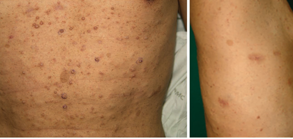
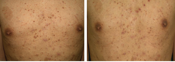
4. Multiple neurofibromatosis: dozens of nodules of various sizes
▶ History of present illness: The patient wanted to remove several relatively large lesions (Figure 1) throughout the body for aesthetic purpose and went through several surgical removal 6-7 lesions at a time under local anesthesia.
▶ Diagnosis: Neurofibromatosis type 1 based on characteristic clinical findings, family history and histological findings.
▶ Treatment: As a nonsurgical treatment of small nodules, selective photothermocoagulation (SPTC) using 1444nm Nd:YAG laser was designed (see Figure 3 and the Discussion below). For larger nodules, less invasive shave excision followed by SPTC with 1444nm Nd:YAG laser was used to treat 10 abdominal lesions. The treated sites are cured without recurrence.

Figure 1. Soft nodules and café au lait spots of various sizes distributed throughout the trunk
[Advertisement] Reandnè Thread Series – Manufacturer: GTG KOREA(www.gtgkorea.com)
Histopathological findings of cutaneous neurofibromatosis
1. Pulpy structure without a membrane covering the tumor, typically located in the dermis infiltrating into the subcutaneous fat layer
2. Multinuclear giant cells are found in the lesion of NF patients. Grenz zone appears between the lesion and the skin.
3. The lesion accompanies fascicles of the thickness of microscopic single cell, which has spherical or fusiform nuclei and indistinct, small cytoplasm. The substrates are faintly dyed and composed of convoluted collagens. Dysplastic neurofibromatosis should be suspected if the cell is dense and atypical. Patients with such lesions are highly likely to show malignancy; they should be periodically observed and the lesion should be surgically removed.

Figure 2. Histology (H&E) of neurofibromatosis. (Left: Intradermal lesion and grenz zone. Center: Cutaneous neurofibromatosis cells. Right: Fascicles)
Discussion on the treatment of cutaneous neurofibromatosis
Neurofibromatosis type 1 (NF-1) is an autosomal dominant hereditary disorder (mutation on the long arm of chromosome 17), characterized by lifelong development of 500-1,000 cutaneous neurofibromatosis originated from the peripheral nerves. Multiple cutaneous neurofibromatosis has a great effect on the patient’s quality of life from aesthetic aspect. Surgical removal is required if the lesion triggers neurological symptoms or is suspected of malignancy. Patients also want surgical removal due to aesthetic reason, but there is still no widely accepted standard for surgical treatment. Surgical resection can be performed under general anesthesia for relatively large lesions, but only limited number of lesions can be removed at a time due to the limit of repeated anesthesia and operational time. Various nonsurgical methods have been attempted for early small and flat nodules in the early stage.
Nonsurgical methods reported by far used electrocautery for resection. This method allows removal of relatively large number of lesions at a single or multiple procedures but is limited by long healing process, scar and discomfort. CO2 laser has been developed later and was attempted for the treatment of cutaneous neurofibromatosis, but it was inefficient because it required longer time for cauterization than eletrocautery. Photocoagulation using Nd:YAG laser was first reported in 2008. The authors reported that Nd:YAG laser for exposed flat lesions resulted in aesthetically superior outcome than existing treatments. They attempted interstitial CO2 laser photocoagulation for large lesions and recommended to use this method for large lesions only because this often results in hypertrophic or atrophic scar. Other recent studies attempted selective resection of neurofibromatosis after differentiating them from normal tissues by confocal laser scanning microscopy, but this method is too complicated and inefficient for small dermatofibroma.

Figure 3. Surgical process: (Left: Shave excision; Center: photocoagulation; Right: After removing Acticoat + Duoderm dressing)

Figure 4. 3 months after the procedure. (Left: before the procedure. Right: After the procedure)
This patient went through several removals under local anesthesia but had difficulties in the process due to anesthesia, lengthy progression after the procedure and scar. I designed a methodology called selective photothermocoagulation (SPTC) combining fat- and water-specific absorption and technical capability of selective coagulation of 1444nm Neodymium: Yttrium Aluminum Garnet (Nd:YAG)LASER(Accusculpt, Lutronic, Korea), the latest lipolysis laser applied to various skin diseases. Such characteristics were found as effective for watery tissues or vascular lesions, and the experience will be published (see reference 4). With this methodology, small and flat lesions were treated by nonsurgical direct photocoagulation, while multiple large, protruding lesions were first excised at the fundus of the protruding tumors by shaving, followed by SPTC for remaining subcutaneous neurofibroma tissues. This method was more efficient at removing neurofibromatosis than existing surgical removal and simple surface laser photocoagulation, induced less recurrence, higher patient satisfaction and superior aesthetic outcome. By using SPTC in multiple cutaneous neurofibromatosis, flat lesions can be removed by nonsurgical direct surface photocoagulation, and larger lesions can be safely and effectively removed on an outpatient setting by shave excision followed by photocoagulation even to the subcutaneous tumor sites.
In conclusion, SPTC using 1444 Nd:YAG laser allows selective procedure because the photocoagulation process can be confirmed with the naked eye, and faster healing due to less heat injury of the surrounding tissues. I recommend this as the most effective minimally invasive procedure at the moment.
References
1. R. R. Anderson, W. Farinelli, H. Laubach et al. Selective photo thermolysis of lipid-rich tissues: a free electron laser study. Lasers Surg Med 2006; 38: 913~919.
2. K. P. Boyd, B. R. Korf, A. Theos. Neurofibromatosis type 1. J Am Acad Dermatol 2009; 61: 1~14; quiz 15~16.
3. T. F. Elwakil, N. A. Samy, M. S. Elbasiouny. Nonexcision treatment of multiple cutaneous neurofibromas by laser photocoagulation. Lasers Med Sci 2008; 23: 301~306.
4. KG Lee, SG Lee, SM Yi, JH Kim, JE Choi, Il-Hwan Kim . Proposing Concept Of Selective Photothermocoagulation (SPTC) and Various Dermatologic Indications By Using 1444nm Nd:YAG LASER. Grapevine, Texas, 31th ASLMS annual conference March 30~April 2, 2011 (Oral Presentation).
5. Koller S, Horn M, Weger W, Massone C, Smolle J, Gerger A. Confocal laser scanning microscopy-guided surgery for neurofibroma. Clin Exp Dermatol. 2009 Dec; 34(8):e670~2. Epub 2009 Jun 22.
6. Elwakil TF, Samy NA, Elbasiouny MS. Non-excision treatment of multiple cutaneous neurofibromas by laser photocoagulation. Lasers Med Sci. 2008 Jul; 23(3):301~6. Epub 2007 Aug 15.
7. Levine SM, Levine E, Taub PJ, Weinberg H.Electrosurgical excision technique for the treatment of multiple cutaneous lesions in neurofibromatosis type I. J Plast Reconstr Aesthet Surg. 2008 Aug;61(8):958-62. Epub 2007 May 25
8. Ostertag JU, Theunissen CC, Neumann HA.
Hypertrophic scars after therapy with CO2 laser for treatment of multiple cutaneous neurofibromas. Dermatol Surg. 2002 Mar; 28(3):296~8.
9. Moreno JC, Mathoret C, Lantieri L, Zeller J, Revuz J, Wolkenstein P. Carbon dioxide laser for removal of multiple cutaneous neurofibromas. Br J Dermatol. 2001 May; 144(5):1096~8
10. Roenigk RK, Ratz JL. CO2 laser treatment of cutaneous neurofibromas. J Dermatol Surg Oncol. 1987
Feb; 13(2):187~90.
- To be continued -
▶ Previous Artlcle : #3. The Most Effective Method for Surgical Treatment of Earlobe Keloid and Prevention of Its Recurrence
▶ Next Artlcle : #5. Removal of Multiple Lipomatosis


