


▶ Previous Artlcle : #2-2. Thermal Effect and Non-Thermal, Non-Cavitational Effect of Ultrasound
Ultrasound cavitation is the formation of microbubbles in a liquid, which gradually become bigger with vibration and explode by ultrasonic energy acting upon the liquid. The dynamics of cavitation bubbles is influenced by the intensity and frequency of ultrasound and physical characteristics of the medium.

[Advertisement] Ultra Skin/Pastelle – Manufacturer: WONTECH(www.wtlaser.com)
The level of cavitation varies; high power cavitation may be strong enough to inflict a serious damage to tissues, while lower power cavitation can only induces a slight change, such as increasing cell membrane permeability. Cavitation starts as a small gas capsule-like cavitation nucleus as small as several nanometers. The bubbles, which work as a cavitation nucleus under the influence of ultrasound field, repeatedly undergoes expansion and contraction. In the process, gases in the surrounding media are gradually accumulated in the bubbles by ‘steady-state diffusion’ during the expansion phase, gradually increasing the size of the bubbles. High-intensity ultrasound would burst the bubbles shortly after an abrupt expansion. Under a standing wave, the sphere-shaped microbubbles would grow and burst symmetrically, and this is called symmetric collapse (Fig 1).
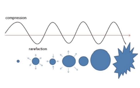
Fig 1. Generation and symmetric collapse of microbubble in Ultrasound field.
Asymmetric collapse occurs when cavitation is formed next to a solid wall, such as skin barrier or blood vessel wall, in which case a high-pressure microjet is generated toward the wall (Fig 2). This high-pressure jet may create a crack or erosion in the wall.
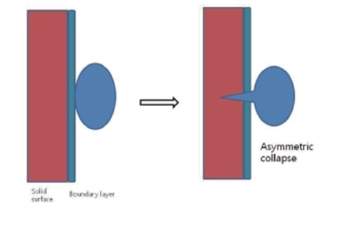
Fig 2. Formation of a jet through acoustic cavitation in a liquid near a surface.
Acoustic microstreaming is a non-thermal action related to the cavitation. When a microbubble repeats expansion and contraction continuously, shearing forces are generated around the bubble, creating a stream of liquid to a direction along the cell wall. This affects the cell structure and cell wall permeability (Fig 3). Cavitation may exert biological effects, therefore, by creating changes in tissues where microbubbles are present. For example, microbubbles can be generated by ultrasound irradiation into the body compartments containing a liquid, such as the kidney, ureter and bladder (e.g., ultrasonic lithotripsy for the treatment of kidney stones). Another example is that a high-intensity ultrasound can create gas bubbles from cavitation nuclei (microbubbles) that are present in the cardiovascular vessel walls. The gas bubbles are mostly created from the ureter or vessel walls and move along the circulation. As mentioned earlier, the presence of gas cavitation and cavitation nuclei is a prerequisite for the formation of bubbles by ultrasound. Bubbles expand and burst rapidly under the influence of high-intensity ultrasound. In human, gas cavitation and microbubbles are commonly detected in the intestines and lungs. Tissues containing these gas cavitations are more prone to vascular damage and hemorrhage. Artificial microbubbles, such as ultrasound contrast agents, can also produce the same effect.
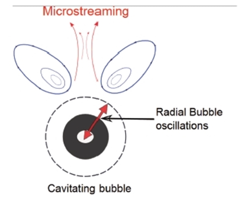
Fig 3. Acoustic micro streaming.
-To be continued-
▶ Next Artlcle : #3-2. Ultrasound Cavitation


