


▶ Previous Artlcle : #11-1. Treatment approaches for Basal Cell Carcinoma of Face in Koreans and Mohs Micrographic Surgery
Treatment
Different surgical methods are applied depending on the location and size of the BCC lesion. BCC occurring in areas with low recurrence such as the chest or limb can be effectively removed by curettage followed by electrocautery, cyrosurgery or traditional excision under local anesthesia, However, the likelihood of scarring and recurrence still remains. Deciding on an optimal treatment requires expertise as it differs depending on the size, infiltration depth, location, and histological form of the lesion. Mohs Micrographic Surgery (MMS) brings the best cure rate and aesthetic results in high-risk areas of the neck, center of the face and around the eyes (H-zone). MMS allows a phased incision of the lesion by examining a frozen section during the surgery and spares the maximum amount of healthy tissues.

[Advertisement] Aileen plus(Long pulsed Nd:YAG Laser) – Manufacturer: FineMEC(www.finemec.net)
The previous incision method removed at least 4mm of the normal tissue without microscopic examination of the 100% of the margins, therefore had a high risk of recurrence. The principles, concepts and stages of the MMS are as follows (Figure 3). Curettage is performed in the center of the lesion to allow microscopic examination of 100% of frozen section margins. The edges are beveled at a 45-degree angle. Preparation of the cross-section of the specimen for the frozen section examination and accurate mapping of cancerous cells with staining are particularly important processes of this procedure.
The stained specimen is placed under 400x microscope to check for remaining cancerous cells, a process which must be carried out by the operating surgeon. In a staged excision, only the area with remaining cancerous cells is removed with minimal excision of the healthy tissues. This way, complete removal of cancer cells as well as the best aesthetic outcome are possible. In Korea, this method has been introduced in university hospitals since the 2000s with limited application. MMS has been covered by national health insurance since a few years ago.
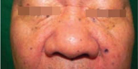
Figure 2. Image of a patient with nevoid BCC syndrome: Early-phase pigmented BCC lesions are scattered around the T-zone of the face in varying sizes; presentation very similar to moles.
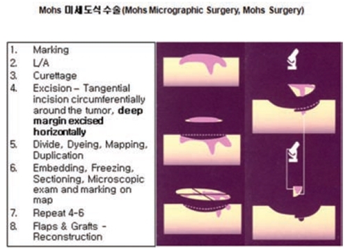
Figure 3. Concepts and stages of Mohs Micrographic Surgery.
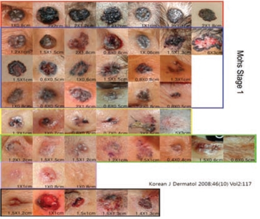
Figure 4. Pigmented BCC is specific to Koreans.
Lesions wth higher degrees of pigmentation and consistency has less subclinical infiltration and can be cured with one stage of Mohs surgery 1(Reference 2).
-To be continued-
▶ Next Artlcle : #11-3. Treatment approaches for Basal Cell Carcinoma of Face in Koreans and Mohs Micrographic Surgery


